Treat Every Arrhythmia
with Personalized Solutions.
Learn From Real-World Cases
![]()
Case Studies

Boston Scientific is developing personalized diagnostic and therapeutic solutions to better understand and treat arrhythmias. See how our personalized portfolio advances electrophysiology for you and your patients.
Share Your Experience
Case Studies
Atrial Fibrillation

Atrial Fibrillation/Left AFL
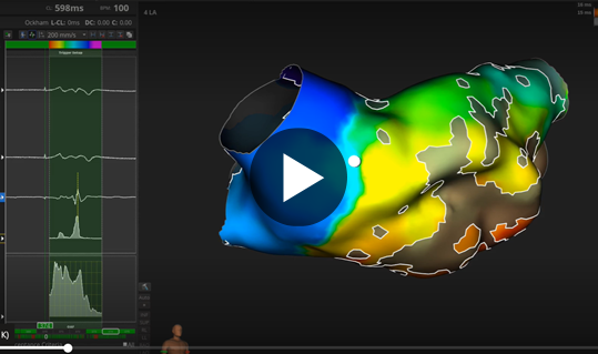
Using DIRECTSENSE™, physicians were able to terminate arrhythmia in a patient who’d presented in atrial fibrillation with durable PVI and fractionated EGMs.
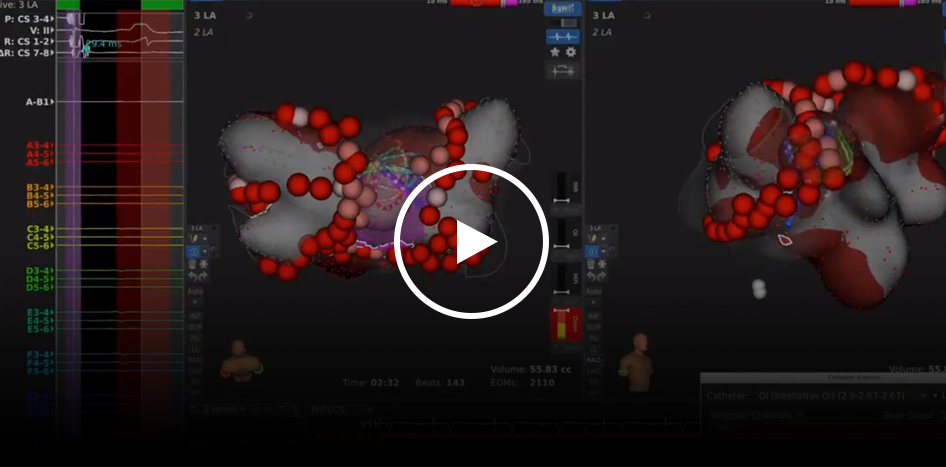
DeNovo Atrial Fibrillation
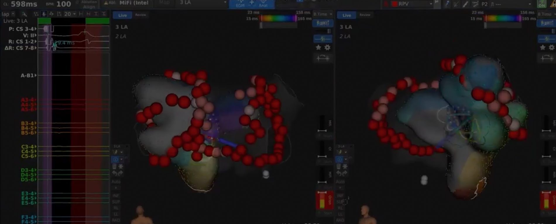
A DeNovo PVI was performed on a PAF patient by first creating a CS-paced map with the INTELLAMAP ORION™ Mapping Catheter with the RHYTHMIA HDx™ Mapping System to assess Left Atrium voltage.
The INTELLANAV MIFI™ OI Catheter with DIRECTSENSE™ Technology was used for ablation with AUTOTAG coloring parameters set to Light Pink <14 Ω, Pink ≥14 Ω, Red ≥17Ω. During PVI, areas of fractionation were highlighted near the RSPV and LIPV using the LUMIPOINT™ Software Module’s complex activation tool.
Fractionated areas were ablated and a floor line was used to anchor them to the LPV and RPV and avoid future LAFL. Validation mapping confirmed PVI and floor line block. The total procedure time was 1:26.
“Local impedance is a very sensitive marker of field response. We notice variability changes when in contact with moving tissue versus more steady. [Local Impedance is a] nice way to tell different tissue properties.”
Video courtesy of Dr. Jose Cuellar-Silva (HCA Houston Healthcare Clear Lake| Webster, Texas)
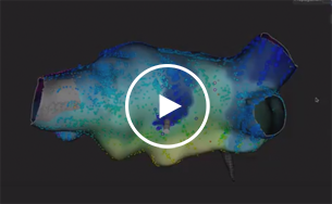
Paroxysmal Atrial Fibrillation
This patient with Paroxysmal Atrial Fibrillation had a second potential in the posterior wall (epicardial connection from CS). The signal was mapped to the roof and ablated. Signals from INTELLANAV MIFI™ OI Catheter begin walking out and disappear.
Video courtesy of Dr. Jose Cuellar-Silva (HCA Houston Healthcare Clear Lake | Webster, Texas)
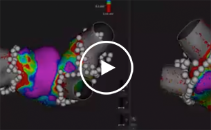
Low Fluoro in AF, CTI Flutter and AT
This case video shows a RHYTHMIA HDxTM low fluoro procedure including a PVI, CTI Flutter and Atrial Tachycardia. Case time was 2.5 hours, with positive outcome and efficiency.
Video courtesy of Dr. Jose Cuellar-Silva (HCA Houston Healthcare Clear Lake | Webster, Texas)
Atrial Tachycardia

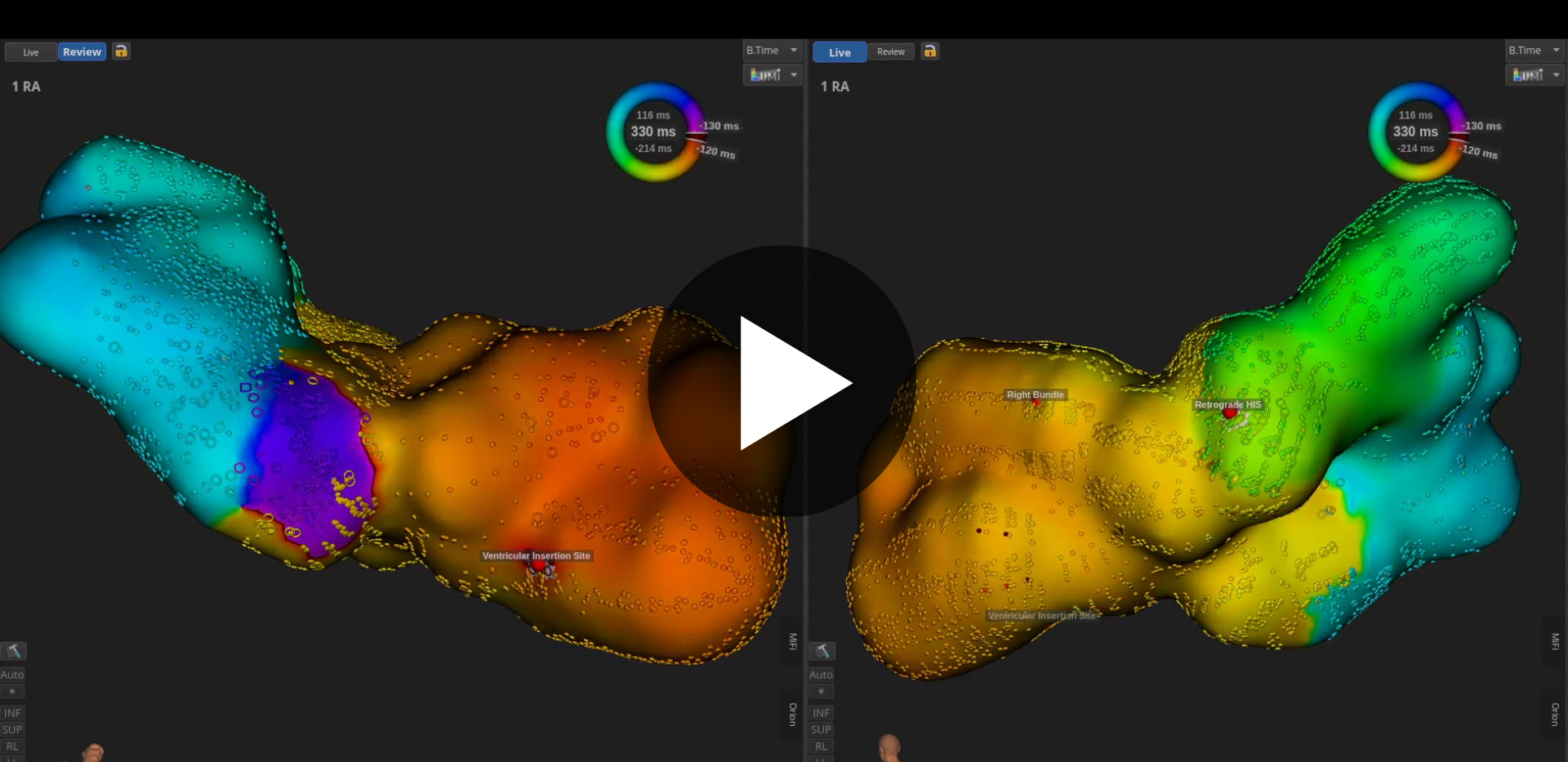
Atrioventricular Reentrant Tachycardia
Physicians using RHYTHMIA HDx™ mapped the entire circuit during tachycardia and identified Mahaim potentials in a patient found to have Mahaim-mediated AVRT.
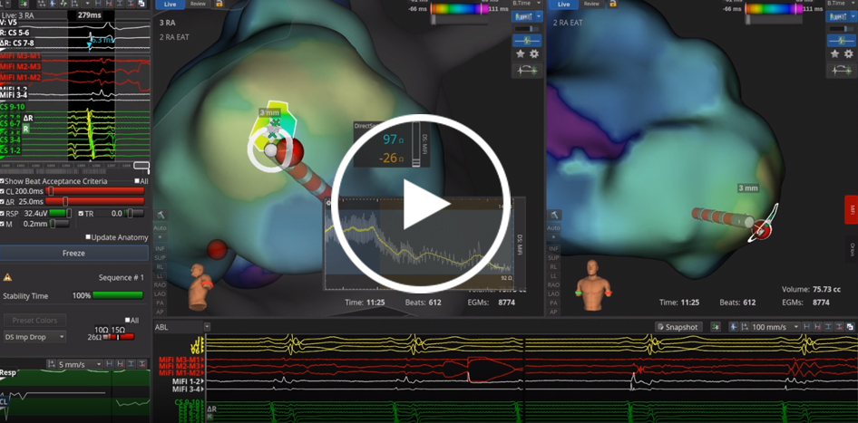
Alternating Tachycardias with 1-Burn Terminations
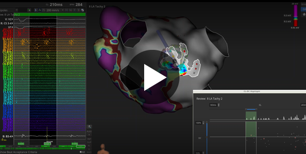
A patient with a complex cardiac history presented with recurrent atrial flutter and atrial tachycardia. Dr. Sunil Sinha sequentially mapped and terminated alternating tachycardias with RHYTHMIA HDx™ in less than 36 minutes. Using LUMIPOINT™ and DIRECTSENSE™, both tachycardias were terminated with a single RF lesion each.
Video courtesy of Dr. Sunil Sinha (Johns Hopkins Heart and Vascular Institute|Baltimore, Maryland)
Focal AT on CTI
A focal atrial tachycardia was localized to the CTI. RHYTHMIA HDx™ and LUMIPOINT™ were used to differentiate between typical atrial flutter and focal AT on the isthmus and to target ablation. The tachycardia was fully terminated within a few seconds of RF delivery.
Video courtesy of Dr. Jose Cuellar-Silva (HCA Houston Healthcare Clear Lake |Webster, Texas)
Macroreentrant Circuit
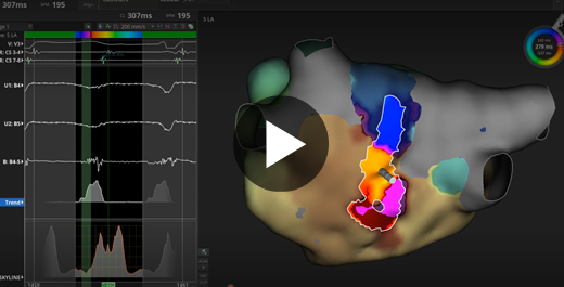
A 66-year-old male, with previous PVI and CTI, presented in tachycardia. When the INTELLAMAP ORION™ Mapping Catheter was close to the LAA/LSPV ridge, the tachycardia terminated. The tachycardia was reinduced and the LA map was completed. Long, fractionated EGMs covering a majority of the cycle length were found at the ridge anterior to the LSPV. LUMIPOINT™ Software Module’s SKYLINE™ tool confirmed a macroreentrant circuit at the ridge, corresponding with the fractionated EGMs. After approximately 1.5 seconds of ablation, the tachycardia was terminated.
Video courtesy of Dr. Bruce Hook (Lahey Hospital & Medical Center | Burlington, Massachusetts)
Atrial Flutter

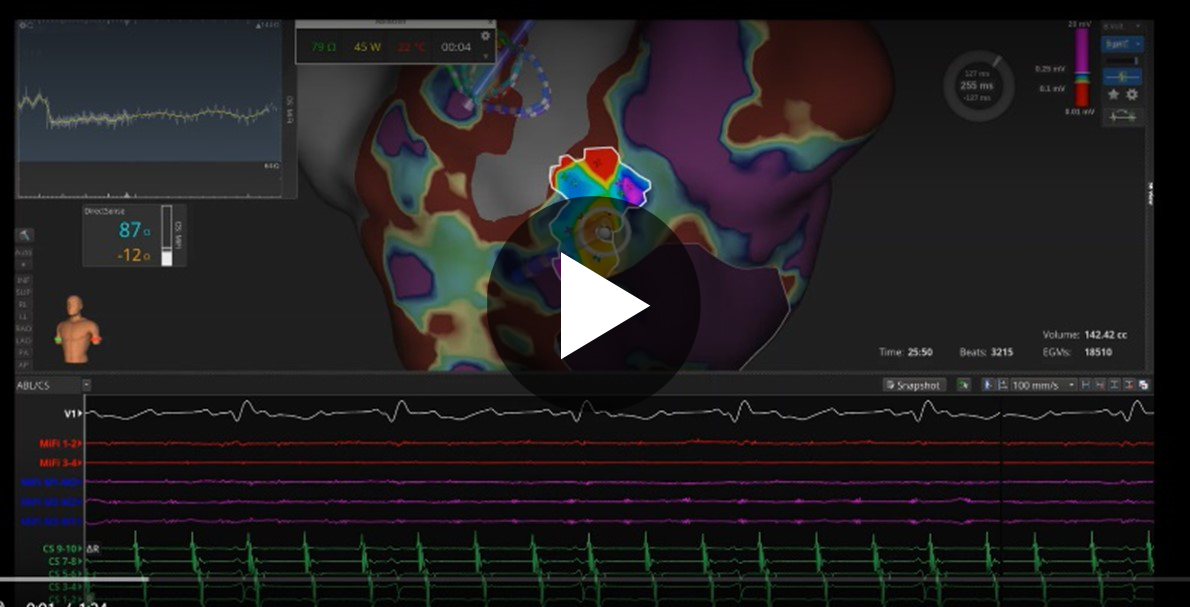
Mitral Annular-Dependent AFL
With RHYTHMIA HDx™, physicians collected low-voltage signals and identified the mitral annular-dependent driving circuit in a patient with atrial flutter.
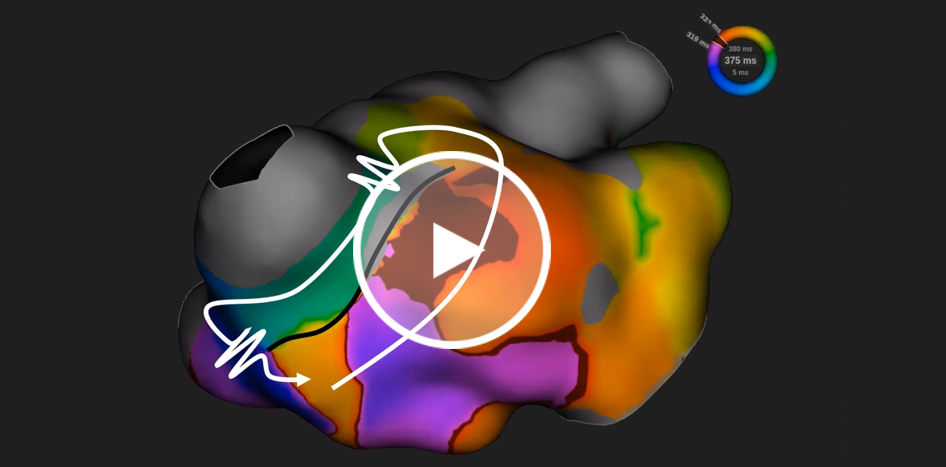
LAFL Originating from RSPV
Following typical left atrial flutter ablation, an atypical flutter was mapped in the LA using INTELLAMAP ORION™. The RHYTHMIA HDx™ Mapping System identified and annotated low-voltage signals in the RSPV and mapped the entire cycle length. The first lesion terminated AFL in 5 seconds and subsequent lesions reisolated.
Video courtesy of Dr. Patrick Whalen (Wake Forest Baptist Health|Winston-Salem, North Carolina)
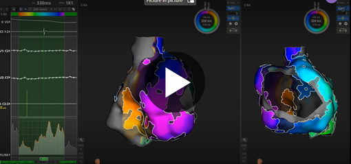
Atypical Right Atrial Flutter: Critical Isthmus Identification
In this patient case, physicians using RHYTHMIA HDx mapping and LUMIPOINT™ identified the circuit’s critical isthmus, enabling termination of AFL.
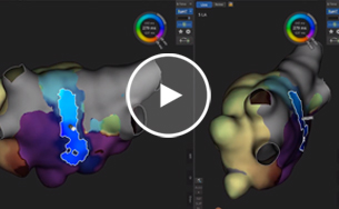
Roof-Dependent LAFL
Dr. Cuellar-Silva identified a roof-dependent left atrial flutter enabled by a gap in a previous posterior wall isolation. Voltage on the posterior wall was <0.02mV. LUMIPOINT™ rapidly highlighted the best target for ablation. LAFL was terminated within 3 seconds of RF delivery.
Video courtesy of Dr. Jose Cuellar-Silva (HCA Houston Healthcare Clear Lake |Webster, Texas)
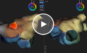
Biatrial Flutter
A complex left atrial flutter mapped appears to use the RA to jump over previous anterior line and complete the circuit. LUMIPOINT™ helped determine a precise location for an efficient single-burn termination.
Video courtesy of Dr. Jose Cuellar-Silva (HCA Houston Healthcare Clear Lake |Webster, Texas)
Ventricular Tachycardia

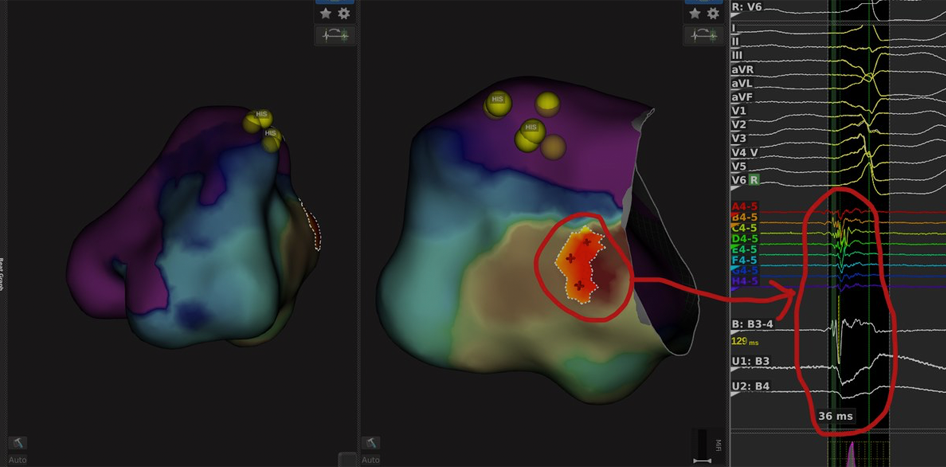
Ventricular Tachycardia (PVC)
A patient presented experiencing a PVC every ~6th beat. Mapping the RV with the INTELLAMAP ORION™ Mapping Catheter, the RHYTHMIA HDx™ Mapping System and using the LUMIPOINT™ Software Module’s SKYLINE™ tool assisted with rapid identification of the true earliest activation at the basal mid-septal region.
At the site of earliest activation, DIRECTSENSE™ Technology was used to monitor stability and the micro-fidelity electrodes on the INTELLANAV MIFI™ OI Catheter allowed visualization of the fractionated signals during a PVC.
DIRECTSENSE Technology was used to guide ablation with AUTOTAG color parameters set to Light Pink <15 Ω, Pink ≥ 15 Ω, Red ≥ 20 Ω. The PVCs were terminated following ablation of the fractionated signals.
Image courtesy of Dr. Jason Chinitz (Southside Hospital | Bay Shore, New York)
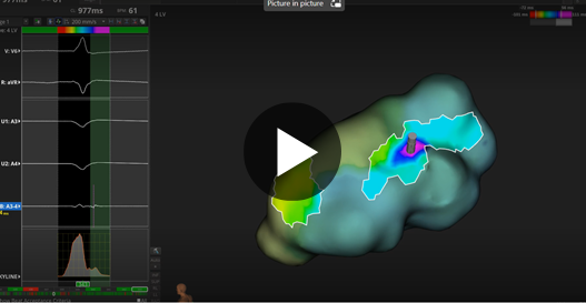
VT Late Potential Identification
In this procedure, a patient with ventricular tachycardia underwent cardiac mapping with RHYTHMIA HDx. In 15 seconds, LUMIPOINT™ Software Module highlighted two areas with abnormal late potentials.
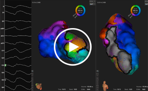
Ventricular Tachycardia
Patient with a history of ischemic cardiomyopathy was treated for recurrent ventricular tachycardia. Dr. Gallagher utilized the RHYTHMIA HDx™, INTELLAMAP ORION™, and LUMIPOINT™ software module to map the entire tachycardia circuit. Local Impedance guided ablation with INTELLANAV MIFI™ OI terminated VT with a linear ablation through the critical isthmus. AUTOTAG demonstrate red tags with >15ohm drops.
Image courtesy of Dr. Peter Gallagher (Nebraska Heart Institute, Lincoln, Nebraska)
