
Advance to
Direct Visualization
Cholangioscopy Image Reference Guide
The intent of this reference guide is to help familiarize physicians with the appearance of biliary findings. Images of normal bile, cystic and hepatic ducts are presented to illustrate and familiarize the user with the appearance of a healthy, normal ducts and to contrast with the appearance of the biliary tree in a variety of pathologic states. Images featuring various types of benign and malignant conditions are represented in this guide, including strictures, villous lesions, stone disease, and more.
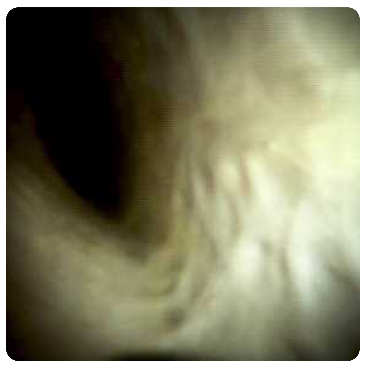
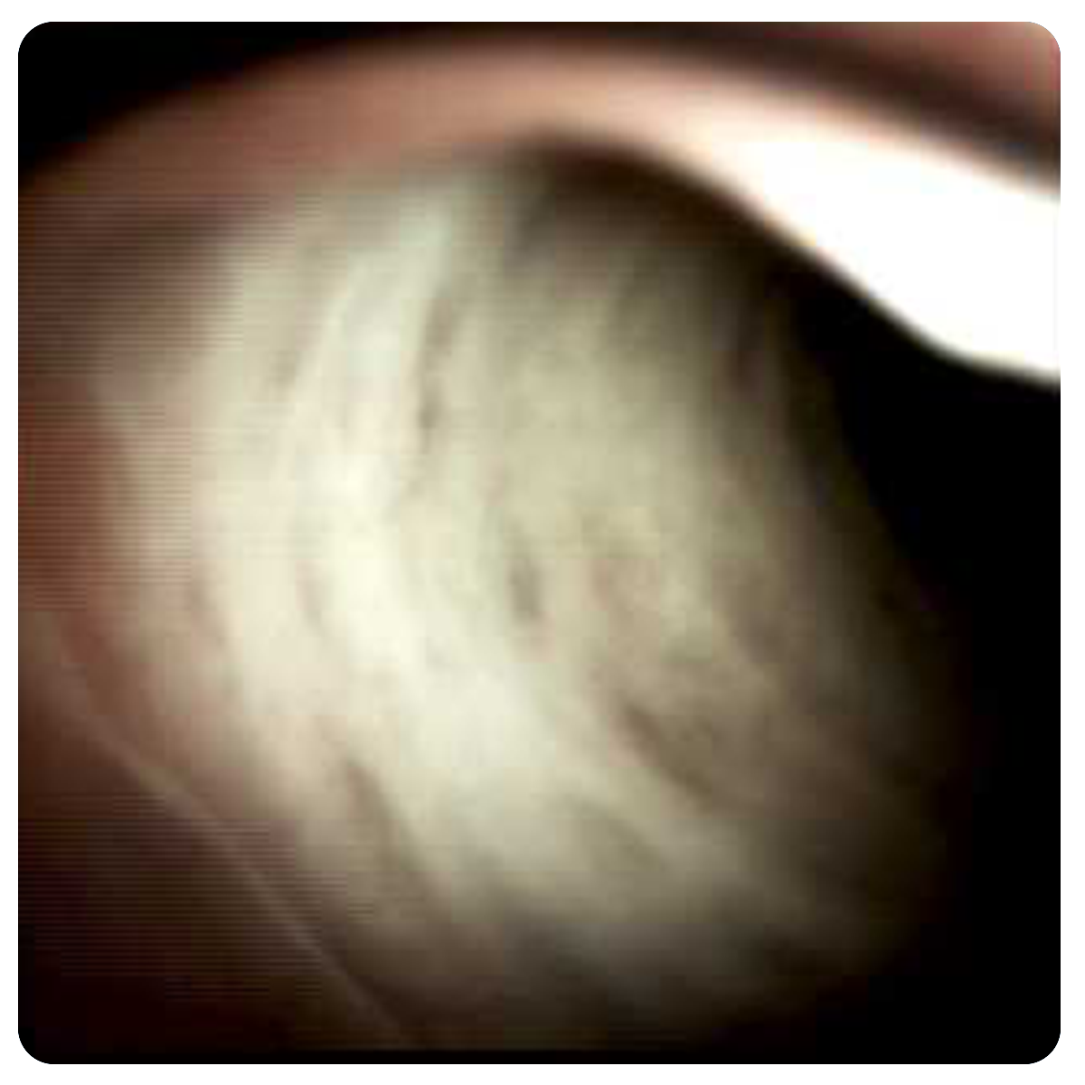
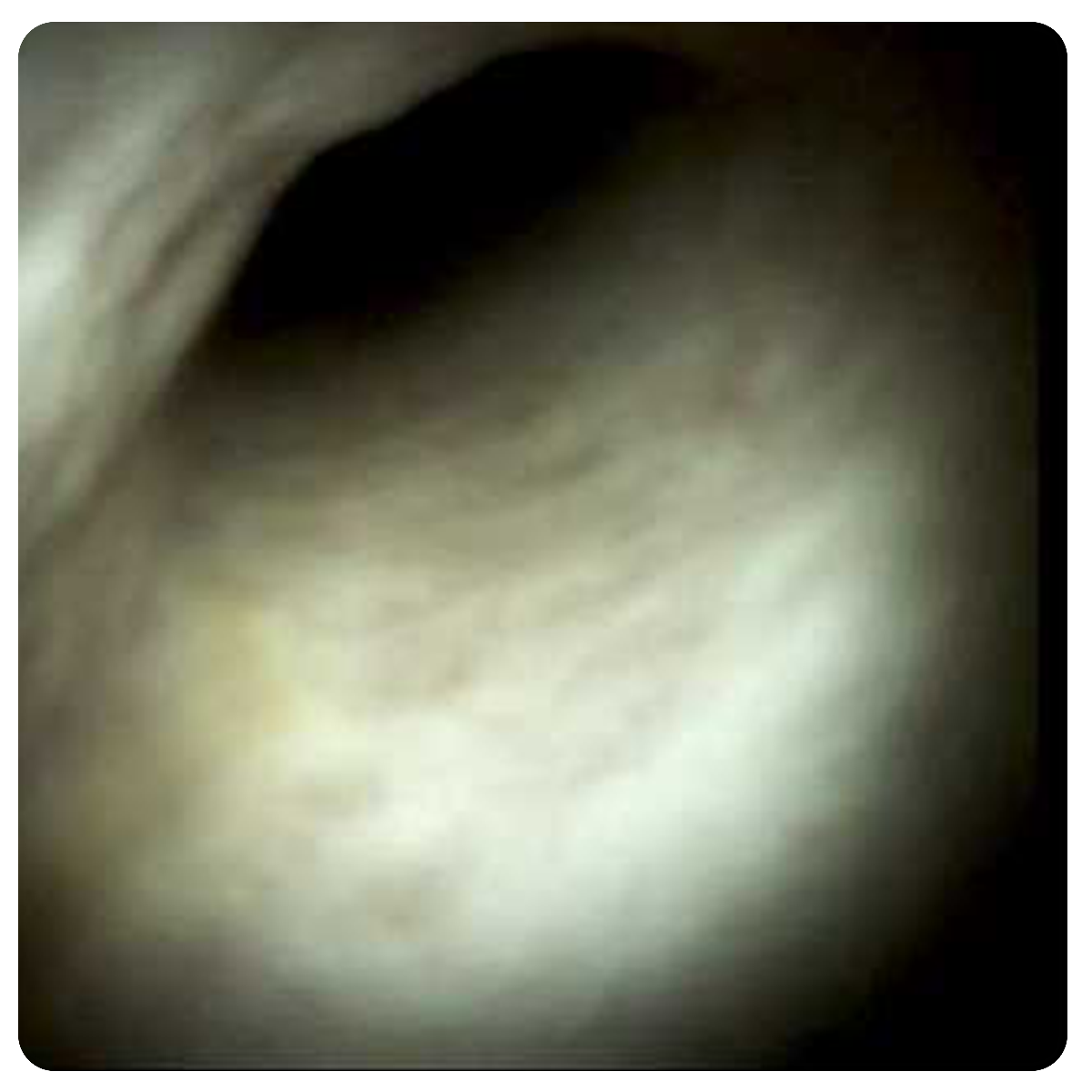
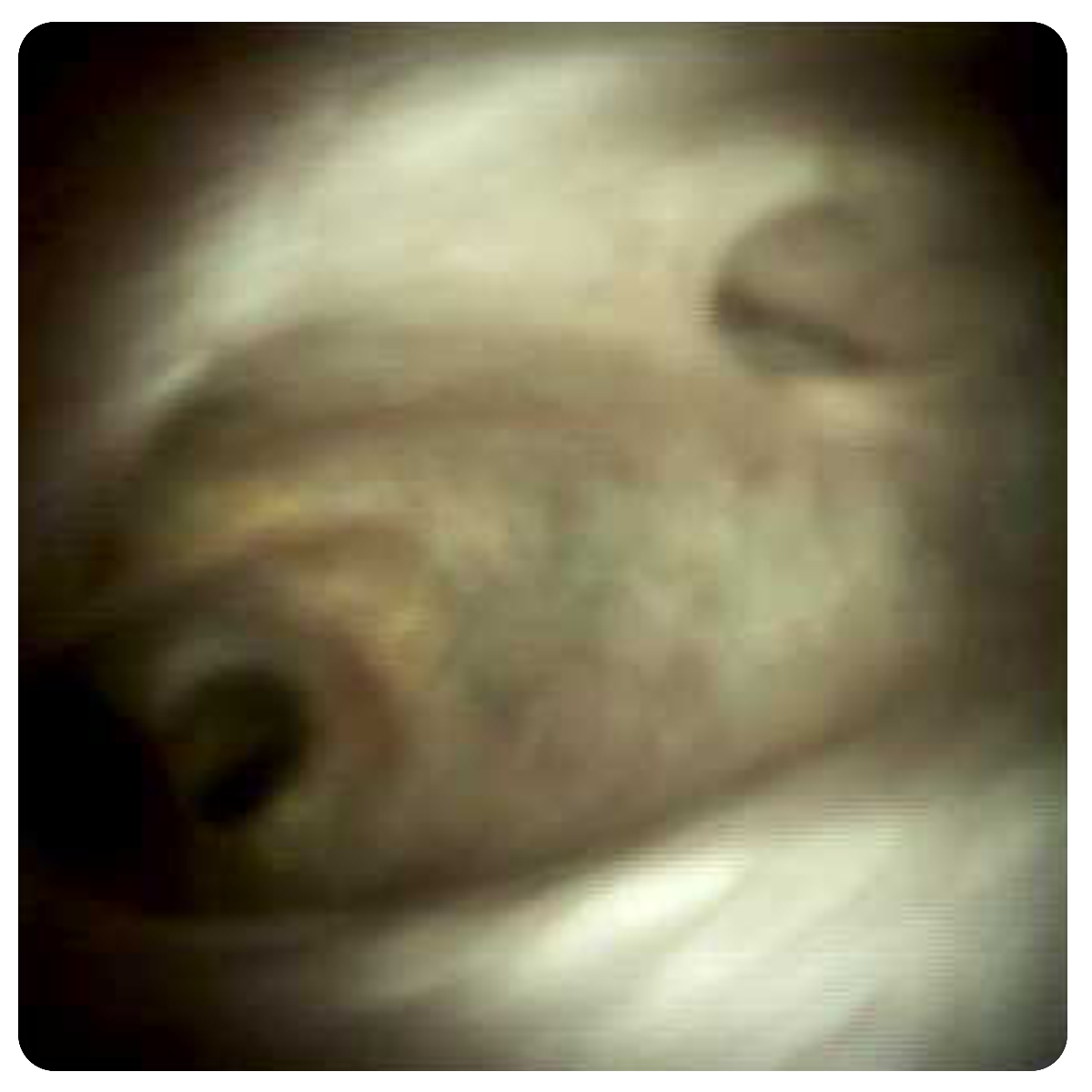
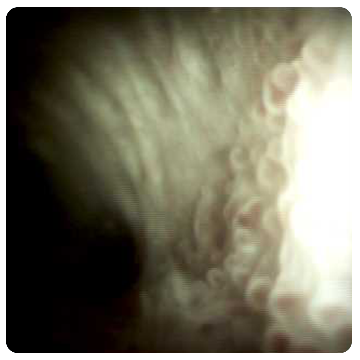
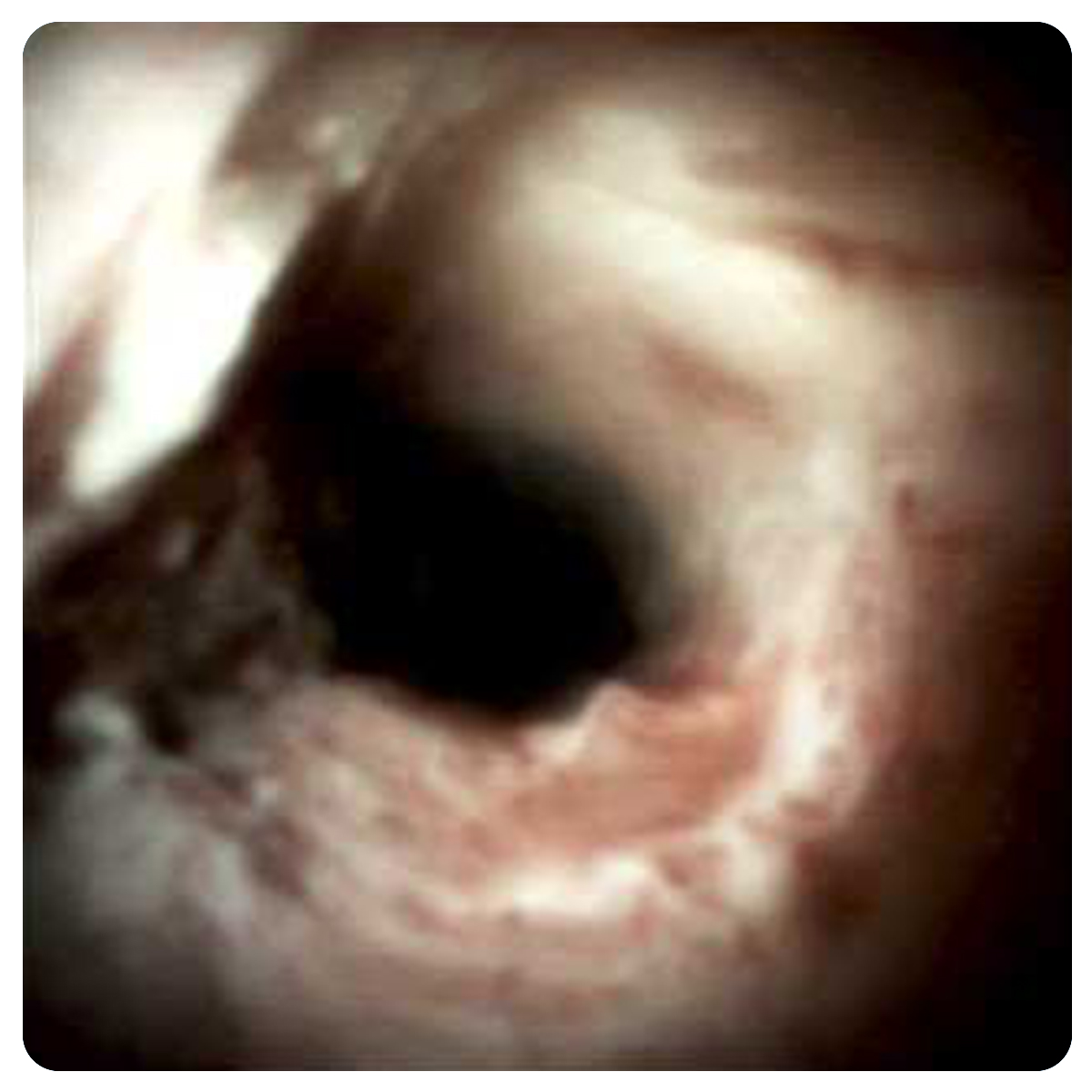
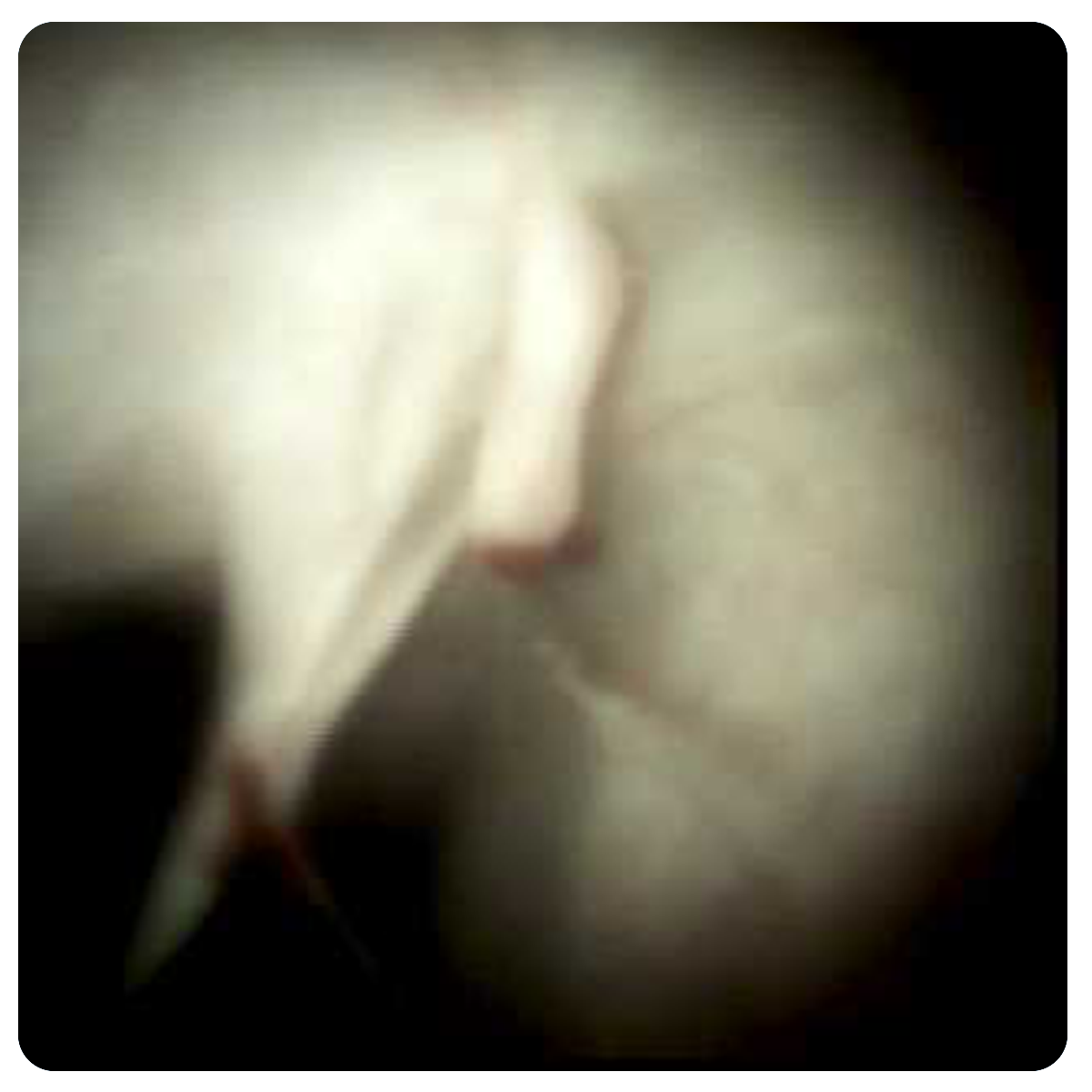
Normal DuctsThroughout the biliary tree, the mucosa has a soft creamy pearly color. Mucosal depressions can be seen which become more apparent in the distal bile duct. They tend to disappear during luminal distention. The vascular pattern is well defined, the vessel contour is crisp and the vascular network is evident. Villous formation is also more noticeable in the distal bile duct. It tends to be uniform, avascular and of short stalk. |
Mucosal Depressions
Shallow Depressions

Normal Vascular Pattern

Normal Villous Pattern

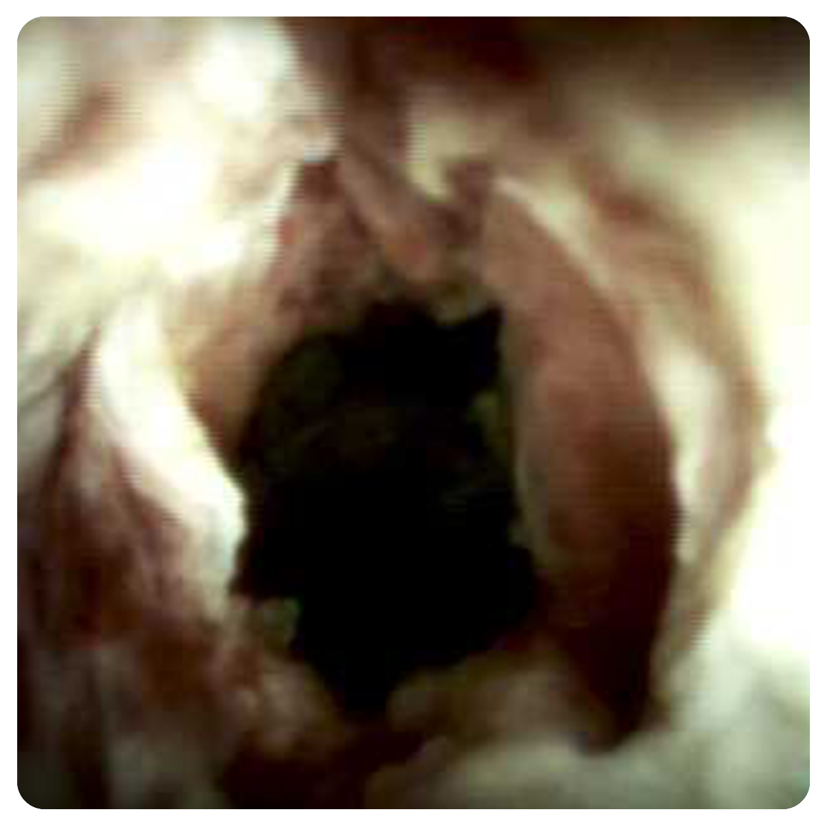
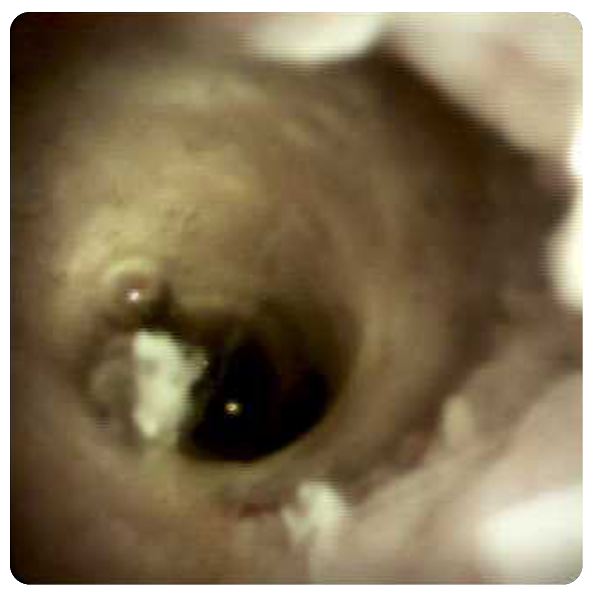
Normal Common Bile Duct

Normal Cystic Duct Common Bile Duct Junction

Normal Common Hepatic Duct

Normal Deep Left Hepatic Duct

Normal Deep Right Hepatic Duct

Normal Left Hepatic Duct Hilum
Normal Mucosa

Normal Papillary Area

Normal Right Hepatic Duct Hilum

Normal Right Hepatic Duct
Normal Suprapapillary
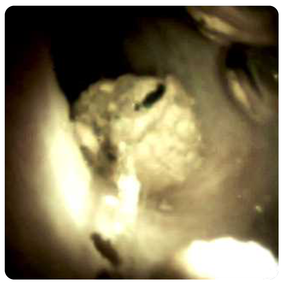
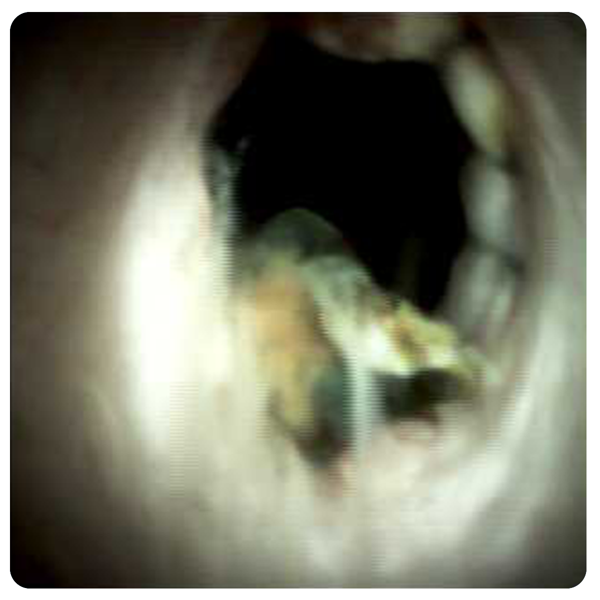
Cystic Duct Stone
Intrahepatic Stone
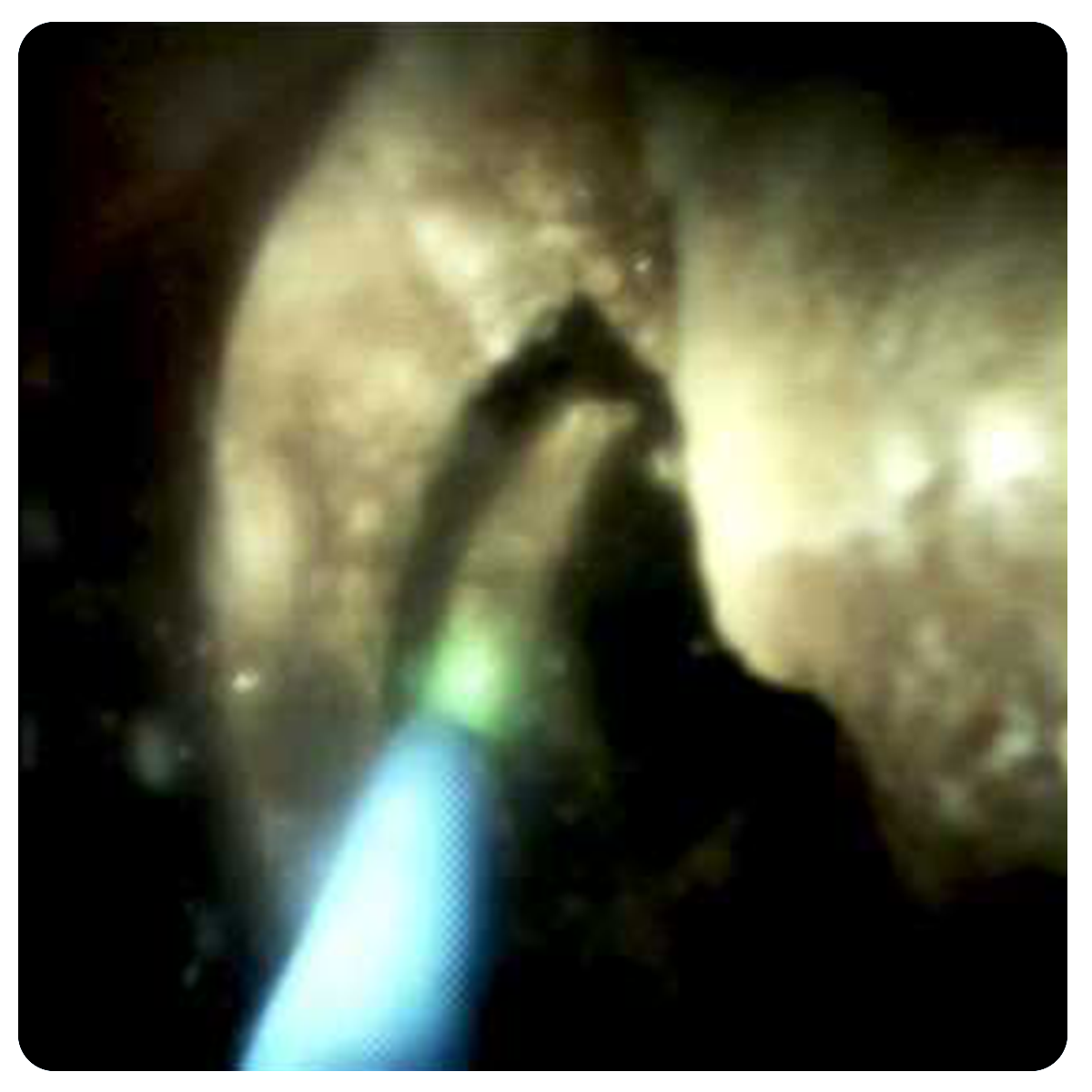
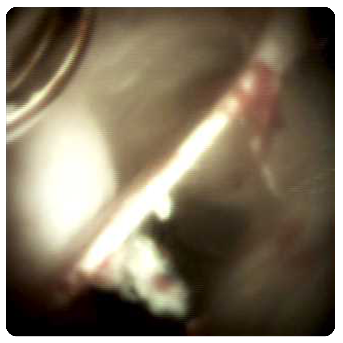
Homium Laser Lithotripsy
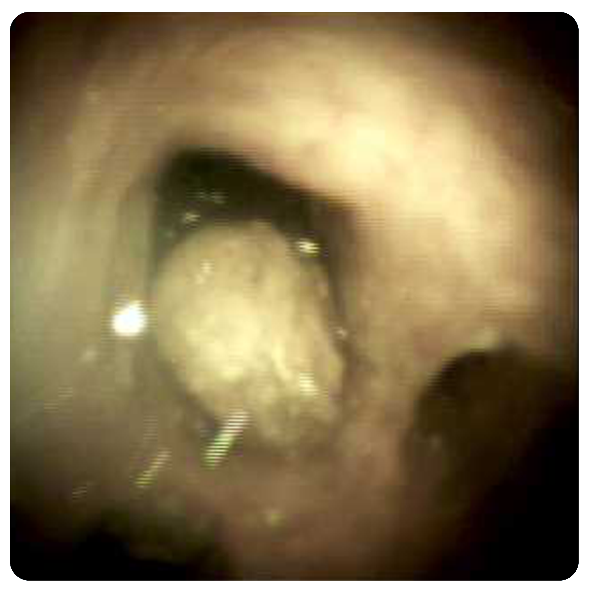
Villiform Formation
Villous Lession

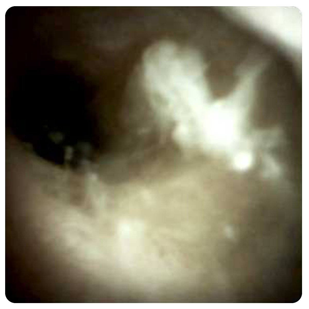
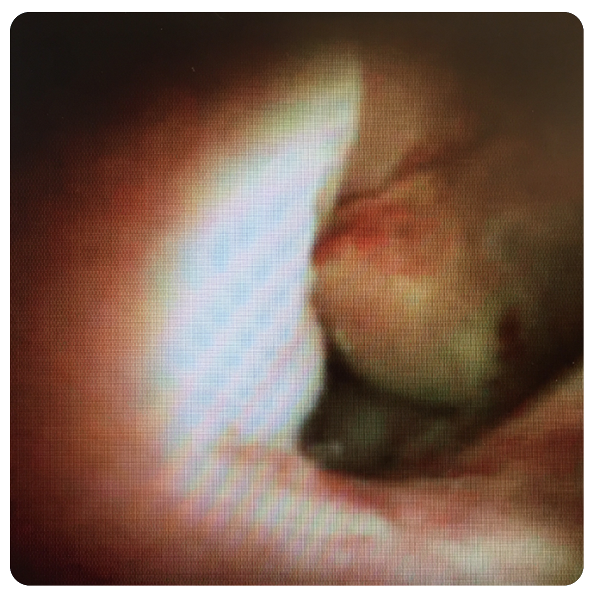
Exophytic Vascular Tissue
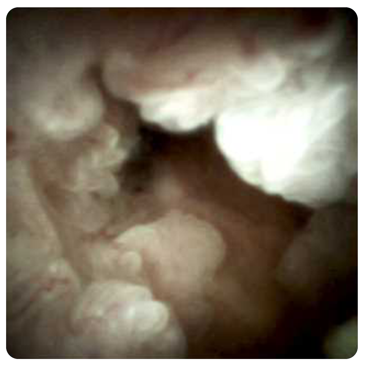
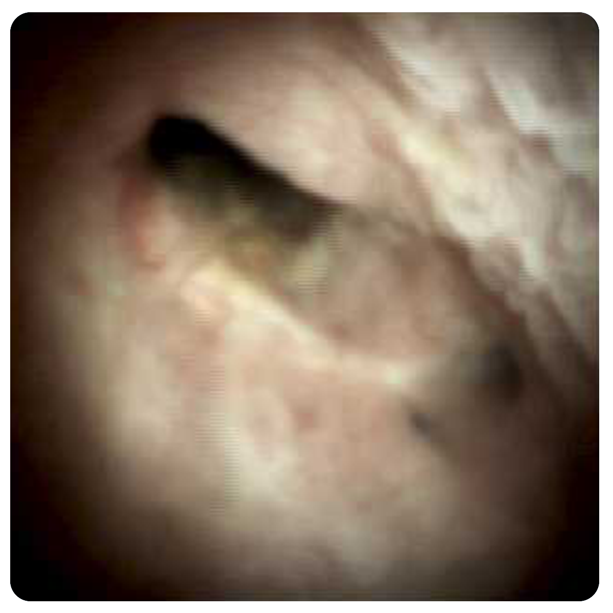
Characteristics of a Malignant Stricture
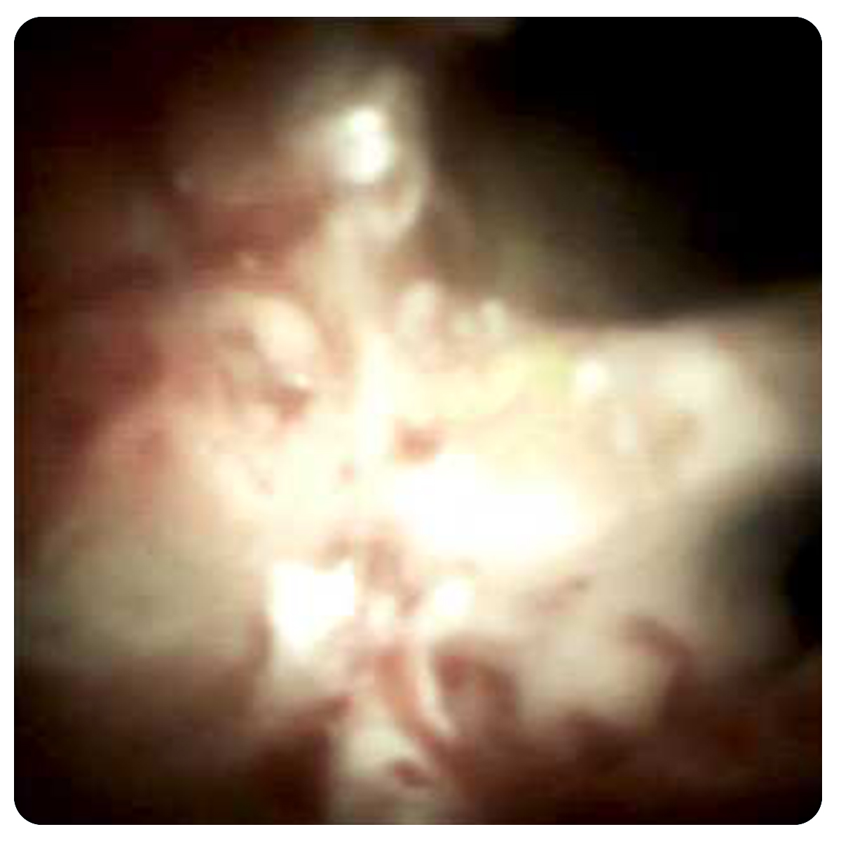
Cholangiocarcinoma Extension
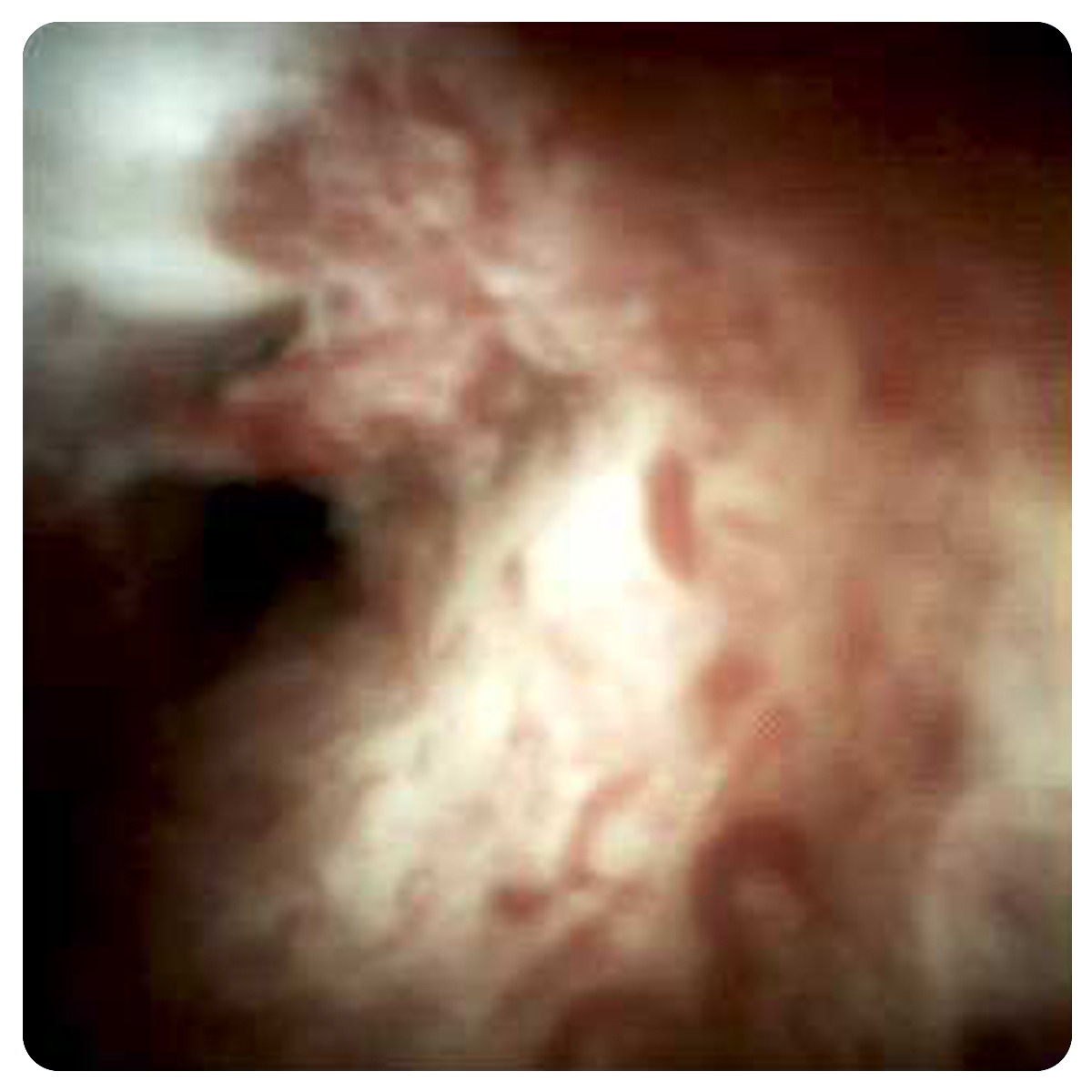
Increased Vascularity
Cholangiocarcinoma STX
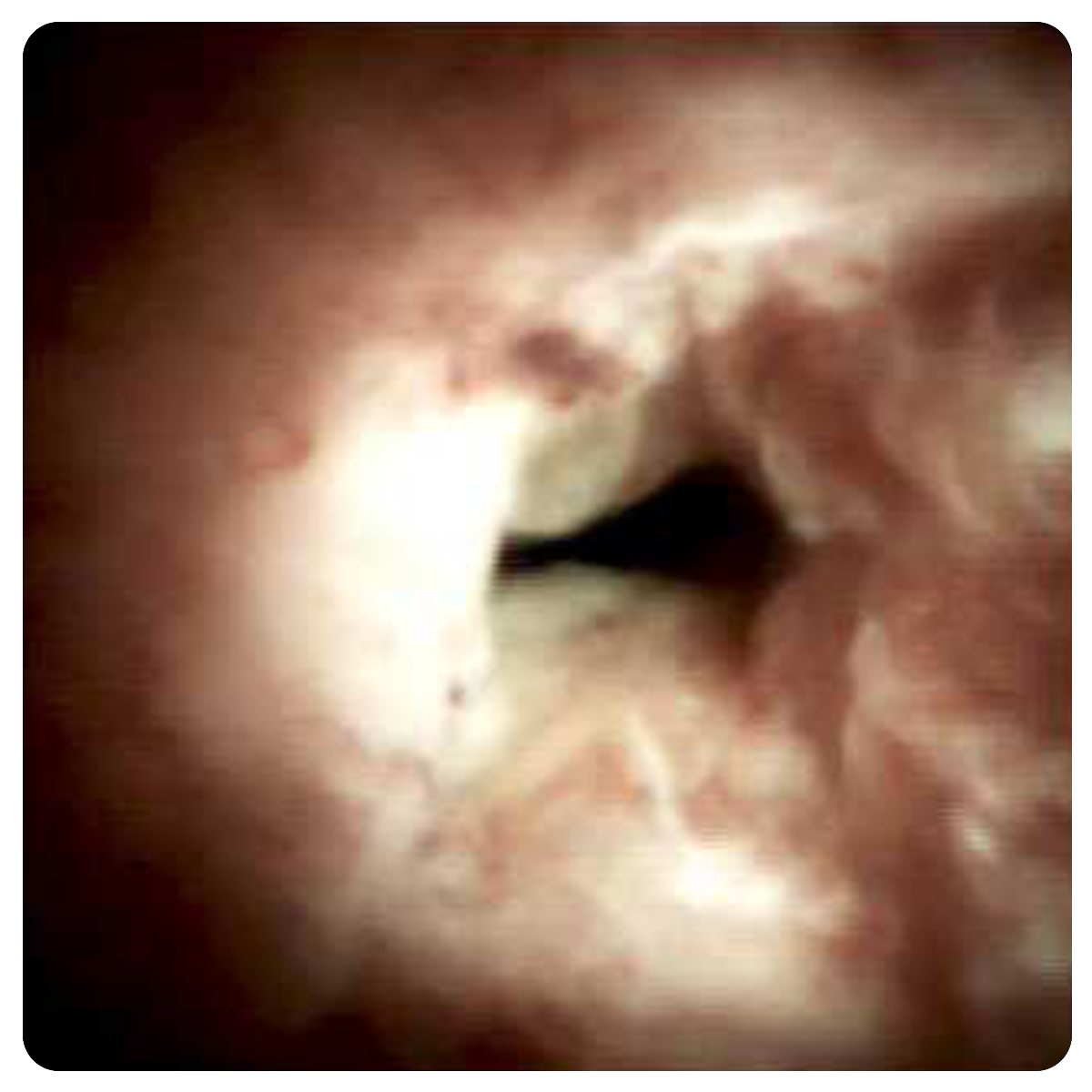
Vascular Lakes

Filling Defects Showing Surgical Staples
Desquamated Epithelium II

Filling Defects Showing Surgical Staples II
Stone Debris
Desquamated Epithelium


Biliary Anastamosis Four Years
Biliary Anastamosis Two Years
Biliary Anastamosis Six Months

Native Cystic Duct Biliary Anastamosis Four Years

Biliary Anastamosis 18 Months Intrahepatic Stones
Epithelial Bridge
Epithelial Bridge II

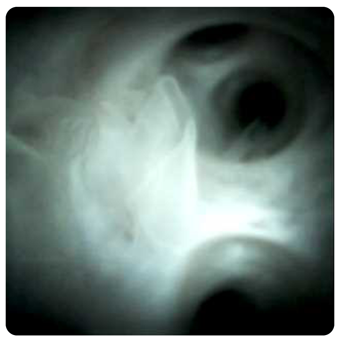
Portal Vein Thrombosis Portal Cholangiopathy

Portal Vein Thrombosis Portal Cholangiopathy IV

Portal Vein Thrombosis Portal Cholangiopathy II

Portal Vein Thrombosis Portal Cholangiopathy III
Hemobilia
ACCESS EDUCARE
SpyGlass Discover Digital Catheter customers
Watch: Making the Case for Percutaneous Cholangioscopy
![]()
Stay up to date
Sign up to receive periodic emails about SpyGlass Discover Digital Catheter, as well as our commitment to sustainability and clinical education, and more.
![]()
Connect with a rep
Request a rep to learn about the SpyGlass Discover Digital Catheter system.