Obturator branch (corona mortis variant) embolization
Courtesy of Dr. Christopher Grilli I ChristianaCare
Presentation
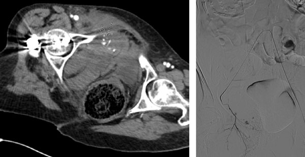
90-year-old female presented to the emergency room as a trauma alert after a fall from standing. Pertinent PMH of atrial fibrillation on Eliquis. Patient reporting abdominal and right hip pain. Noted to be hypotensive upon arrival with systolic pressures in the 70s. Massive transfusion protocol (MTP) initiated. CT of the abdomen and pelvis obtained demonstrating a comminuted fracture of the right superior pubic ramus with active arterial bleeding noted in the adjacent pelvic musculature (CT image 1, arrows). IR consulted for angiogram with possible embolization.
Intervention
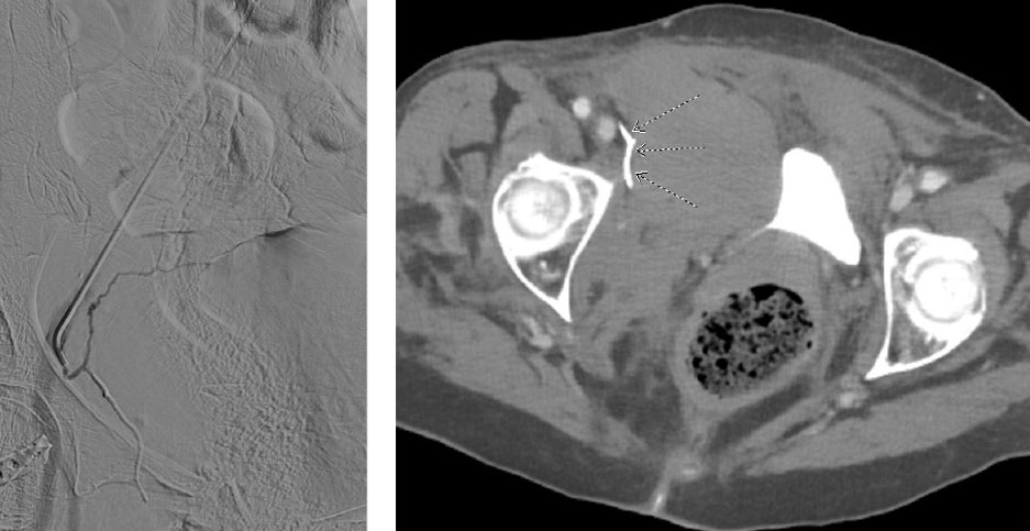
Arterial access obtained via the left common femoral artery. A Berenstein catheter utilized to select for the contralateral right common and subsequently external iliac arteries where arteriography was performed. Variant arterial anatomy was noted with the obturator artery arising from the inferior epigastric (corona mortis). The inferior epigastric and replaced obturator were selected with a 2.4 F Direxion™ Torqueable Microcatheter and Fathom™ Steerable Guidewire where arteriography demonstrated multiple pseudoaneurysms and active extravasation arising from the replaced obturator artery. Embolization was performed with 0.1 mL of Obsidio Embolic. The microcatheter was removed and post embolization arteriography demonstrated occlusion of the obturator without nontarget embolization.
Outcome
The patient’s BP stabilized in the room immediately post embolization. Post-op there were no additional signs of bleeding with stable hgb and vitals. Follow up CTA was performed 2 days post embolization with no further sign of extravasation or pseudoaneurysm. The cast of Obsidio Embolic is noted in the follow up CTA (CT image 2, arrows). The patient was discharged to a SNF 4 days post intervention.


Results from case studies are not necessarily predictive of results in other cases. Results in other cases may vary.
