Boston Scientific accounts are for healthcare professionals only.

OPAL HDx™ Mapping System
Configure or select a product to continue to order
- Overview
- Clinical data
- Technical specifications
- Ordering information
- Training
- Service plan
- Resources
- OPTIMAP™ Integration
How it works
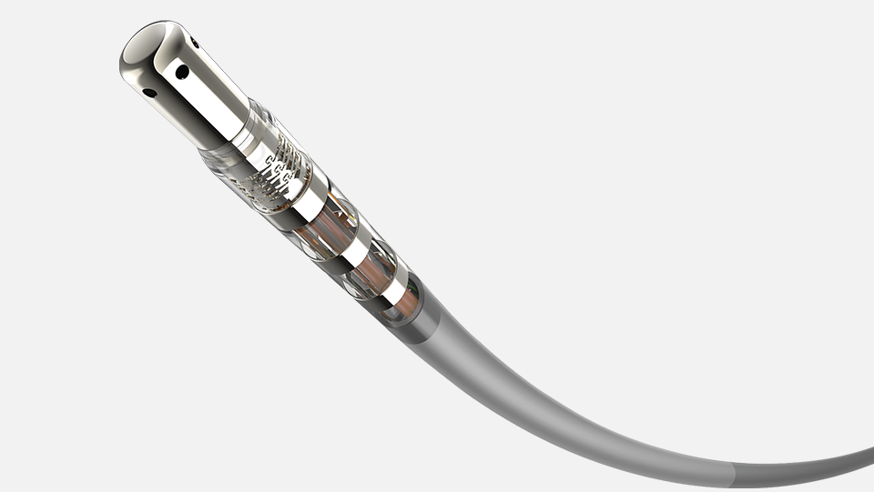
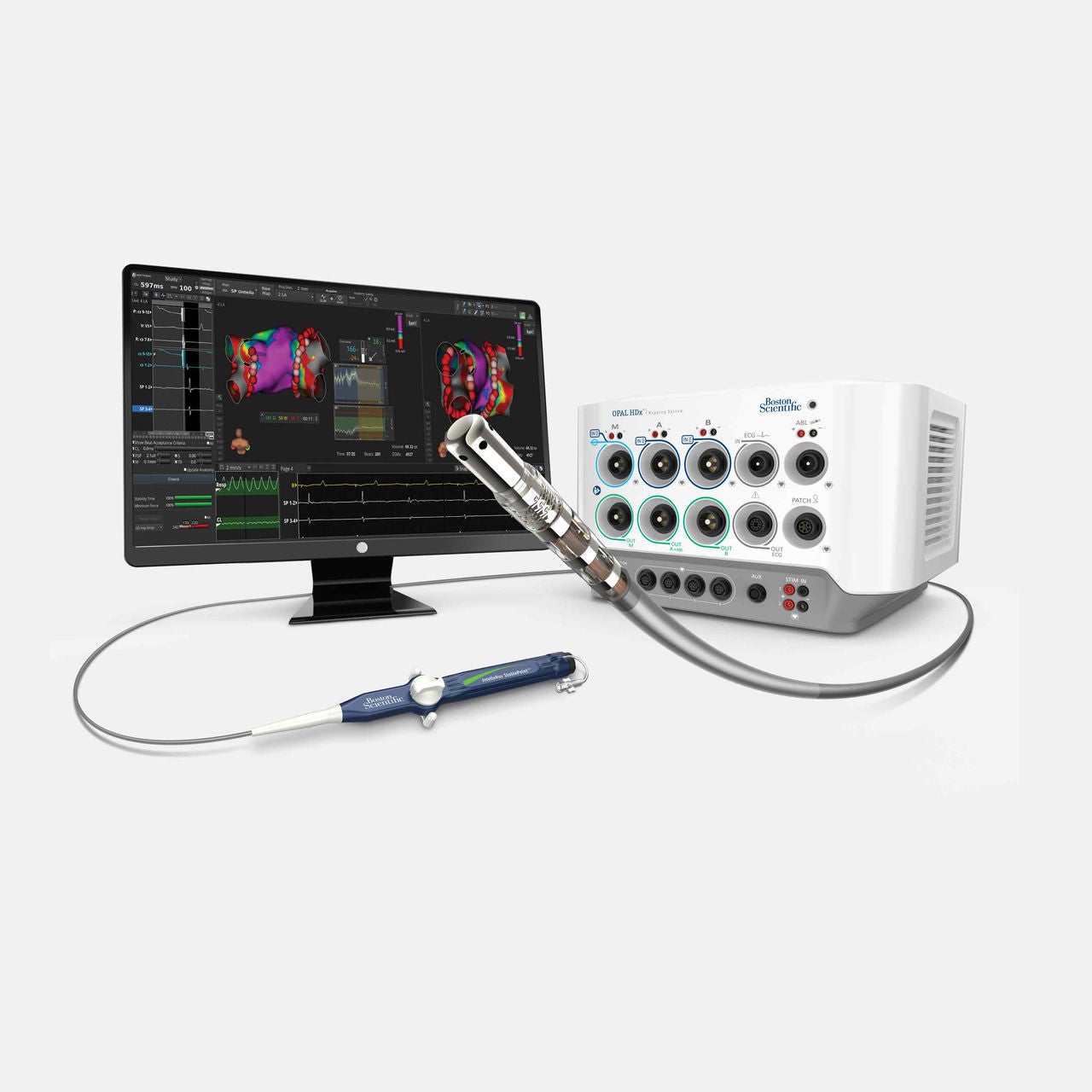
The OPAL HDx™ Mapping System is optimized for FARAPULSE™ PFA and integrates with a full suite of catheters and software modules, combining real-time validation, and advanced mapping to support workflows from pulmonary vein isolation (PVI) to complex arrhythmias.
The mapping solution optimized for FARAPULSE™ PFA
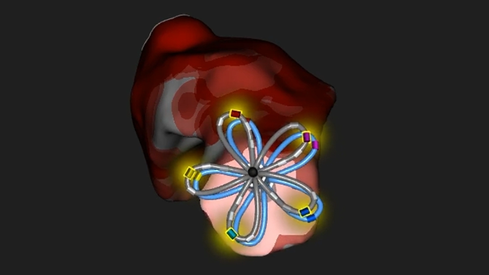
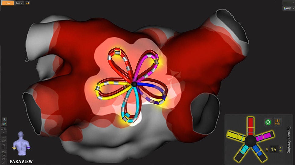
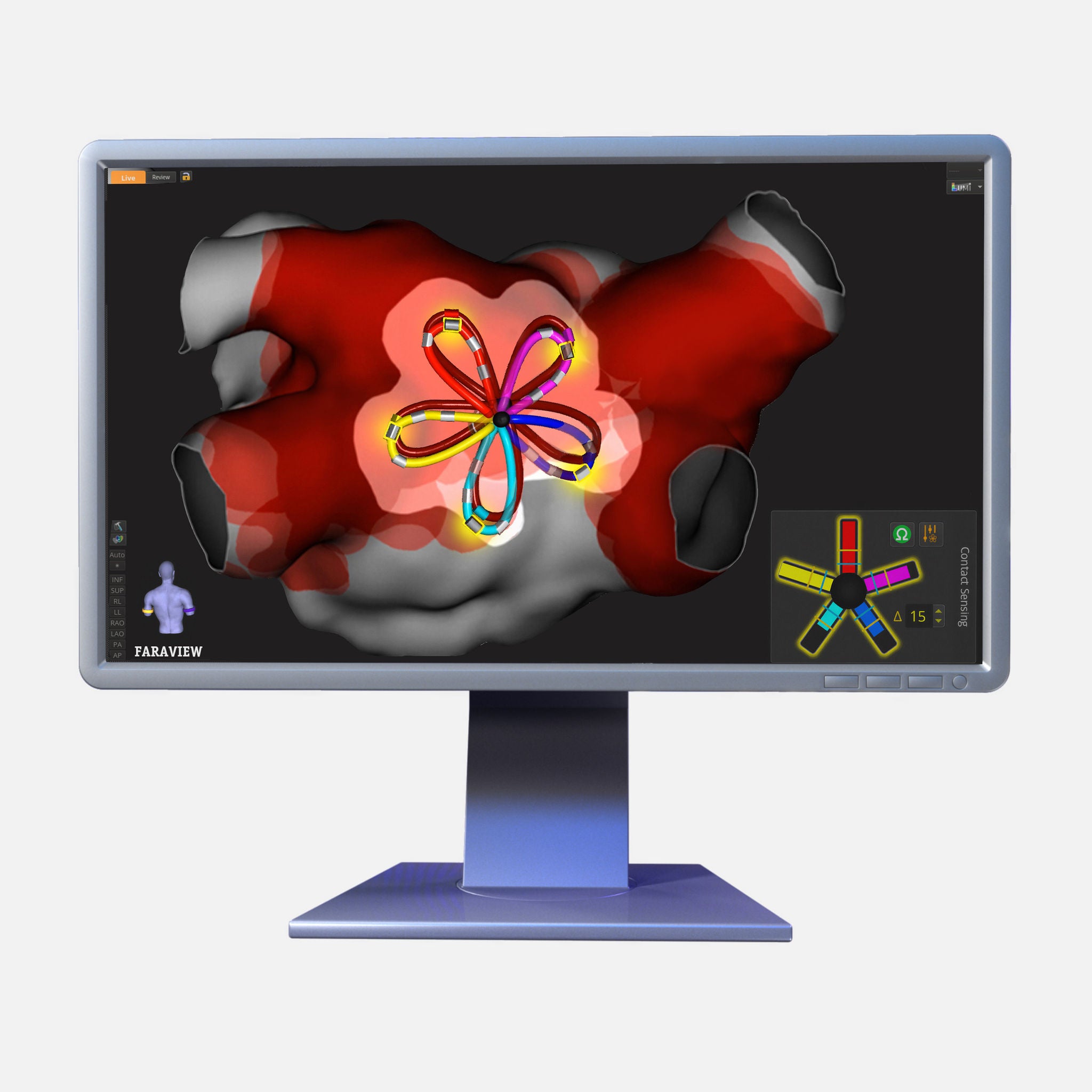
The FARAVIEW™ Software Module provides the optimal experience for FARAPULSE™ PFA procedures, unlocking workflow simplicity and procedural confidence. FARAVIEW provides procedural feedback to enable physicians to clearly see catheter position, catheter contact, and estimated PFA field coverage before and after energy is delivered.

Advanced mapping tools
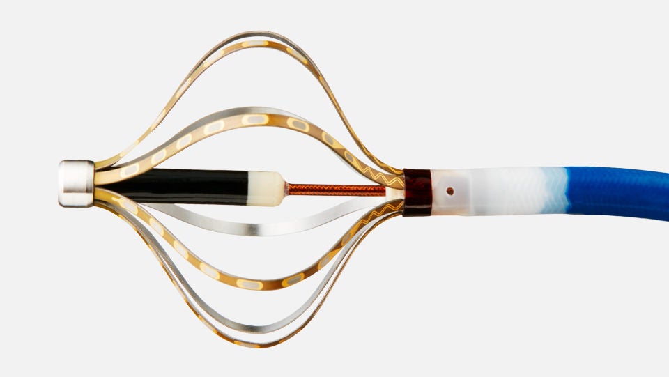
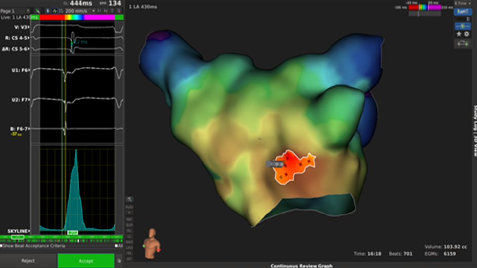
The OPAL HDx Mapping System integrates high-density mapping, intelligent annotation, and advanced electrogram analysis algorithms to localize even the most complex arrhythmias with precision and confidence.
Future mapping innovations
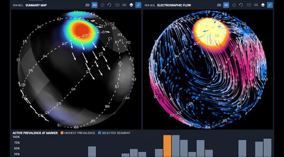
Boston Scientific is continuously advancing the OPAL HDx system through frequent, innovative updates that expand capability and performance. The OPAL HDx system will be furthering capabilities through integration with OPTIMAP™, introducing electrographic flow mapping to support the identification of nonpulmonary vein sources of atrial fibrillation.
The OPTIMAP™ System is 510(k) cleared and is not yet available for sale in the United States.
Clinical data
DISRUPT-AF real-world registry2
Safe, efficient single-catheter map and ablate workflow with the FARAWAVE™ NAV Pulsed Field Ablation (PFA) Catheter, visualized with FARAVIEW™ Software Module on the OPAL HDx Mapping System. The DISRUPT-AF real-world registry (n=477) showed a 0.2% catheter-related complication event rate, predictable procedures and an average fluoroscopy time of 3.8 minutes.
Sub-analysis: FARAWAVE NAV with OPAL HDx vs. non-nav enabled or competitive systems
Evidence from the DISRUPT-AF sub-analysis and other independent clinical investigations highlights the performance of the FARAWAVE NAV Catheter with the OPAL HDx Mapping System. Together, these analyses demonstrate procedural efficiency advantages.3,4 Separately, chronic remapping studies showed consistently high rates of pulmonary vein isolation (PVI) and posterior wall ablation (PWA) durability.5,6
NAVIGATE-PF trial7
The NAVIGATE-PF clinical trial was a multi-center, first-in-human feasibility study that assessed the acute and chronic accuracy of FIELDTAG™ Technology on the FARAVIEW™ Software Module in visualizing pulsed field energy delivery by the FARAWAVE NAV catheter. The study included two phases:
Phase I (acute assessment):
Phase 1 included 30 patients that underwent PV isolation, with 30% also receiving posterior wall ablation.
PFA markers were placed during PFA application and high-quality, post-ablation HD maps with INTELLAMAP ORION™ Mapping Catheter were completed in 15 pts. PFA markers were overlaid on the HD map and contours were drawn at the outer border of the low voltage areas (defined as ≤0.5mV) where at least 2 overlapping PFA markers were dropped.
Phase II (chronic assessment):
Included 20 patients at 1 center to evaluate chronic lesion durability and alignment between acute PFA tag locations and chronic low voltage borders.
NAVIGATE-PF8: 60 day remap
60-day remapping after FARAWAVE NAV PFA Catheter ablation showed 95% (63/66) PV durability and 86% (12/14) PW durability.
OPAL HDx™ Mapping System
| Feature | Specifications |
| Signal Station | Dimensions (W x D x H): 41.0 x 38.9 x 26.2 (centimeter) 16.2 x 15.3 x 10.3 (inches) |
| Workstation PC | Dimensions Z-series (W x D x H): 17.1 x 47.3 x 44.3 (centimeter) 6.8 x 18.7 x 17.5 (inches) |
| Monitor Display size: 24" | Dimensions (W xD x H): 56.2 x 6.1 x 36.8 (centimeter) 22.1 x 2.4 x 14.5 (inches) |
| Localization Generator | Dimensions (W x D x H): 45.0 x 55.4 x 7.0 (centimeter) 17.8 x 21.9 x 2.8 (inches) |
| System Cart | Dimensions (W x D x H) without monitor: Dimensions (W x D x H) with monitor: |
| Workstation PC Cart Holds the Workstation, monitor, keyboard, and mouse | Dimensions (W x D x H) with monitor: 71.2 x 57.2 x 147.4 (centimeter) 28.0 x 22.5 x 58.0 (inches) |
Ordering information
| UPN Description | UPN# | GTIN# |
| OPAL HDx Mapping Sys Install Bundle 2 EN | M004OPLINSTALL2EN0 | 00191506055370 |
| OPAL HDx Signal Station 2 | M004OPAL10200 | 00191506055325 |
| OPAL HDx WORKSTATION PC | M004OPAL40100 | 00191506055301 |
Reimbursement information
Locate procedural coding and reimbursement information for inpatient, outpatient, office, and ASC settings on our reimbursement page. You’ll also find coding and payment guides for cardiac rhythm management and electrophysiology.
Customer support plans
The ExpertCare Program includes tailored plans offering 24-hour technical assistance and expert collaboration in support of Boston Scientific electrophysiology systems.
Online education
Online medical training and education courses
The EDUCARE online platform makes health care education and training more relevant, more comprehensive, more personal, and more accessible. Register to access a library of procedural videos, case studies, training resources, and events.



The ExpertCare Program is designed to provide you with continued confidence in Boston Scientific products, customer support, and technical services. Technical support is available 24 hours a day. Our service team has the expertise that comes from years of collaboration with our customers to better support your commitment to patient care.
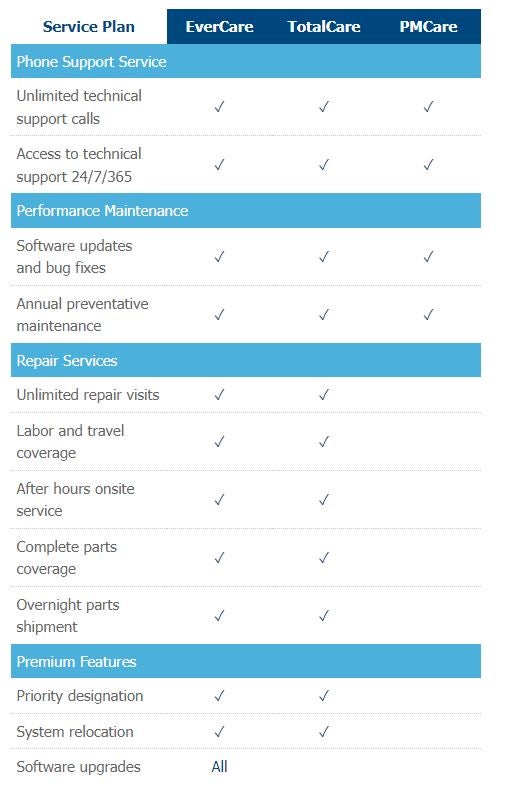
Boston Scientific ExpertCare service plans will provide you extraordinary value in three key areas.
Cost Control - There is nothing worse than an unexpected financial surprise. A prepaid service agreement will eliminate variable maintenance and repairs costs.
Uptime - Time is money and with a prepaid service agreement, you will automatically be in front of the line. Our uptime guarantee is a commitment to delivering the fastest onsite service to systems under an agreement.
Convenience - Prepaid service agreements can eliminate the administrative hassles of purchase orders and associated delays that can slow down the process.
Brochure
Introducing the OPTIMAP™ System
Boston Scientific acquired Cortex Inc., developer of the OPTIMAP™ System, an advanced diagnostic mapping system designed to work in combination with the OPTIMAP™ catheter to visualize atrial fibrillation (AF) wavefront propagation during electrophysiology procedures.
Designed with a proprietary Electrographic Flow (EGF) algorithm, OPTIMAP identifies potential active AF sources: regions where electrical flow repeatedly emanates or diverges. These EGF sources may represent triggers or drivers that contribute to AF and may be difficult to identify using conventional mapping approaches.
Many AF ablation strategies rely primarily on anatomic lesion sets, which may not reflect the functional mechanisms present in an individual patient. OPTIMAP is designed to provide reproducible, visually interpretable outputs and patient-specific source information that may help physicians focus on a more tailored ablation strategy while reducing reliance on empiric ablation.
Future integration with OPAL HDx
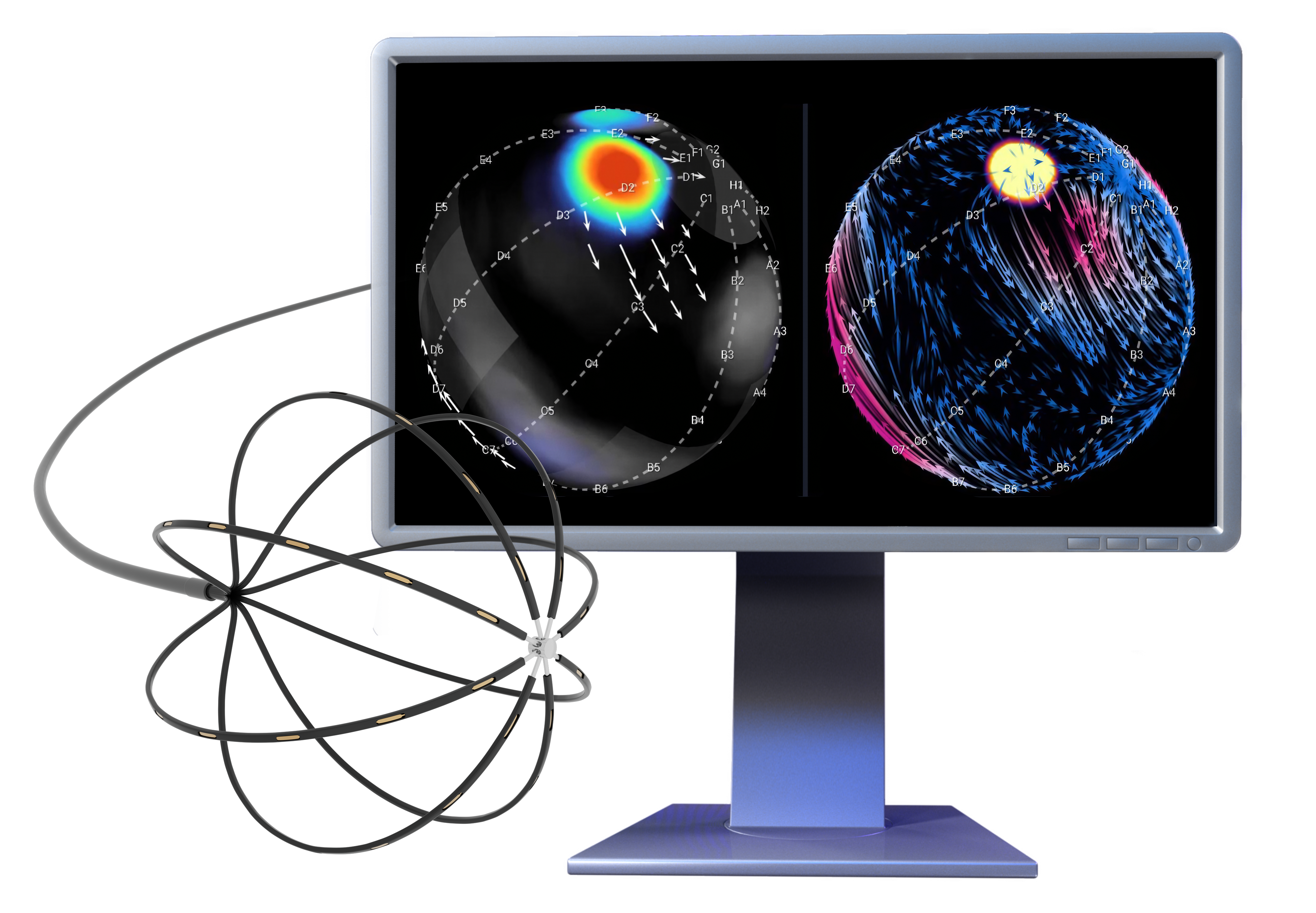
The OPTIMAP system is 510(k) cleared and Boston Scientific is advancing clinical research to further characterize and validate the technology before commercialization. OPTIMAP is planned to integrate with the OPAL HDx mapping system, further strengthening Boston Scientific’s integrated mapping and ablation portfolio to support AF treatment.
Clinical evidence
Early clinical study results demonstrated improved outcomes with pulmonary vein isolation (PVI) plus EGF-guided source ablation compared with PVI-only procedures in redo patients. Ongoing studies aim to reproduce these findings in a larger patient population, and pair EGF mapping with pulsed field ablation (PFA), using OPTIMAP powered by the OPAL HDx mapping system.
Key clinical studies:
- The FLOW-AF study9,11 provided the first randomized evidence for EGF mapping in AF treatment. The study demonstrated a 51% improvement (on an absolute basis) in Freedom from AF at 1 year with PVI plus EGF mapping and ablation versus PVI alone in redo AF patients; p = 0.042. It also showed PVI-only patients with no sources had better outcomes than those with sources: 70% vs. 35%; p = 0.018.
Explore the FLOW-AF study results
- The RESOLVE-AF study10 provided additional evidence supporting EGF mapping in a larger patient cohort. In the redo patient cohort, complete ablation of all active sources following PVI was associated with significantly greater Freedom from AF at 1 year compared with no or partial source ablation: 70% vs. 41%, respectively; p < 0.001.
Explore the RESOLVE-AF redo cohort study results
- The OPTIMIZE randomized controlled trial is designed to evaluate PFA of the pulmonary veins and EGF-identified sources compared with PFA of the pulmonary veins and posterior wall in patients with de novo symptomatic, drug-refractory persistent AF.
- The ReDEFINE-AF randomized controlled trial is investigating whether OPTIMAP-guided extra-PV source ablation using FARAFLEX™ Mapping and PFA Catheter will deliver superior outcomes to an anatomical redo ablation strategy while establishing the clinical value of an integrated Boston Scientific AF workflow on OPAL HDx™.
The OPTIMAP™ System is 510(k) cleared and is not yet available for sale in the United States.
Related products

References:
1. Lepillier A, Maggio R, De Sanctis V, et al. Insight into contact force local impedance technology for predicting effective pulmonary vein isolation. Front. Cardiovasc. Med. 2023 Jul 5;10:1169037. doi: 10.3389/fcvm.2023.1169037.
2. Nair D, et al. Clinical experience with map-and-ablate workflows enabled by the second-generation catheter: findings from the DISRUPT-AF Registry. Presented at AF Symposium 2026.
3. Gabrah, Kirollos, et al. Invasive remapping to assess long-term durability of pulmonary vein isolation for pulsed field ablation with zero fluoroscopy approach. Presented at AHA 2025.
4. Mitacchione, Gianfranco, et al. First clinical experience using a pentaspline pulse field ablation catheter with integrated electroanatomical 3D mapping for pulmonary vein isolation. Heart Rhythm 2025.
5. Rodríguez-Muñoz D, Cobarro Gálvez L, Rajjoub Al-Mahdi EA, et al. Optimized workflow for pentaspline PFA and its impact on PVI durability: Evidence from systematic remapping. Europace. 2026;28(6):euag118. doi:10.1093/europace/euag118
6. Reddy VY, Petru J, Funasako M, et al. Lesion durability of the second generation pentaspline pulsed field ablation catheter NAVIGATE-PF phase 2 results. Circ Arrhythm Electrophysiol. 2026;19(4):e014477. doi:10.1161/CIRCEP.125.014477
7. Reddy VY, et al. Visualization and chronic assessment of electrical field tags for pulsed field ablation of pulmonary veins with the second-generation pentaspline catheter. Presented at HRS 2025.
8. Reddy VY, et al. Chronic lesion prediction of pulsed field ablation using dynamic shape visualization and tags with the second-generation pentaspline catheter. Presented at AF Symposium 2026.
9. Reddy VY, et al. A randomized trial of electrographic flow-guided redo ablation for non-paroxysmal atrial fibrillation (FLOW-AF). JACC: Clinical Electrophysiology. doi.org/10.1016/j.jacep.2024.03.040
10. Boersma LV, Electrographic flow-guided mapping and ablation in redo AF patients: 1-year outcomes of the RESOLVE-AF clinical trial [Poster presentation]. Heart Rhythm Society 2026 Scientific Sessions.
11. Verma A, et al. PVI-only is not enough for all patients with persistent AF: A FLOW-AF subgroup analysis. Heart Rhythm, 22(5), 1170–1178. doi.org/10.1016/j.hrthm.2024.10.037