










Conventional imaging and endoscopic retrograde cholangiopancreatography (ERCP) alone often lack the sensitivity and precision needed for confident diagnosis and management of indeterminate biliary strictures and difficult stones.
At Boston Scientific, our evidence-backed cholangioscopy portfolio allows direct, high-definition visualisation of ductal anatomy to guide targeted tissue sampling and the treatment of complex stones.
Our broad range of single‑use scopes is designed to provide precise procedural control, supporting confident access and intervention across biliary and pancreatic procedures. Engineered to adapt to varied and complex anatomy, the portfolio helps promote consistent performance and efficient workflows.
Proven Performance in Cholangioscopy
We are advancing cholangioscopy-guided care with our SpyGlass™ DS II Direct Visualization System, which has been adopted in more than 85 countries, potentially impacting over 250,000 procedures since 2015.1
Our legacy of innovation in this space spans more than two decades:
- 20 years since FDA clearance of the first Single-Operator Cholangioscopy System SpyGlass™
- 2015: CE Mark obtained for the SpyGlass™ DS System
- 2021: CE Mark obtained for the SpyGlass™ Discover System
- 2024: Long‑term strategic collaboration established with Scivita Medical
The platform reflects our global commitment to offering minimally invasive technologies that support first-time procedural success.
Evidence Supporting the Use of Cholangioscopy During ERCP
Studies demonstrate the value of incorporating biliary cholangioscopy into ERCP, with benefits extending to both patients and healthcare providers.
34%
of patients out of 118 experienced a change in surgical plan when the SpyGlass™ DS System was used to map malignant involvement in the biliary of pancreatic ducts before surgery.²
85%
of patients who underwent diagnostic ERCP with cholangioscopy had their clinical management altered, in a clinical study of 298 patients.³
89%
stone clearance after a single ERCP session may allow patients to avoid repeat endoscopic interventions or more invasive procedures for difficult biliary and pancreatic stones.4
Expanding Imaging Capabilities in Cholangioscopy
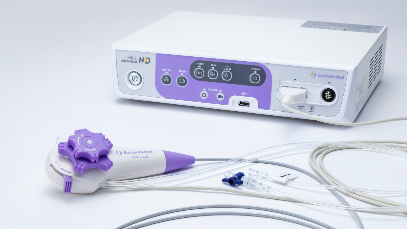
Our recent collaboration with Scivita Medical enabled us to co‑develop imaging solutions to meet evolving needs. The Scivita Medical Single-Use Cholangioscopy System offers broad functional versatility, with expanded size configurations and the ability to capture still endoscopic images and record video.
The platform also supports:



Image‑adjustment features further enhance visualisation control, supporting efficient documentation and real‑time decision‑making during procedures.
Cholangioscopy for Stone Management
Electrohydraulic Lithotripsy (EHL) for First-Time Procedural Success
When conventional balloon or basket extraction is insufficient, whether due to stone location, size, specific stone features, or patient anatomy, cholangioscopy-assisted lithotripsy with EHL is recommended by the European Society of Gastrointestinal Endoscopy (ESGE) as a therapeutic option for difficult bile duct stones.*2
A 2021 systematic review and meta-analysis supports EHL as an effective, evidence-backed approach to achieving first-time ductal clearance.5
95%
stone fragmentation success rate5
82%
single-session fragmentation and duct clearance5
4.6%
adverse event rate5
Clear Residual Bile Duct Stones
The Autolith™ Touch Biliary EHL System is designed to provide improved patient safety, treatment efficacy, and ease of operation while achieving stone clearance.
When combined with the SpyGlass™ DS System, it has been shown to enable a high stone clearance success rate in single session treatment.6
It includes:
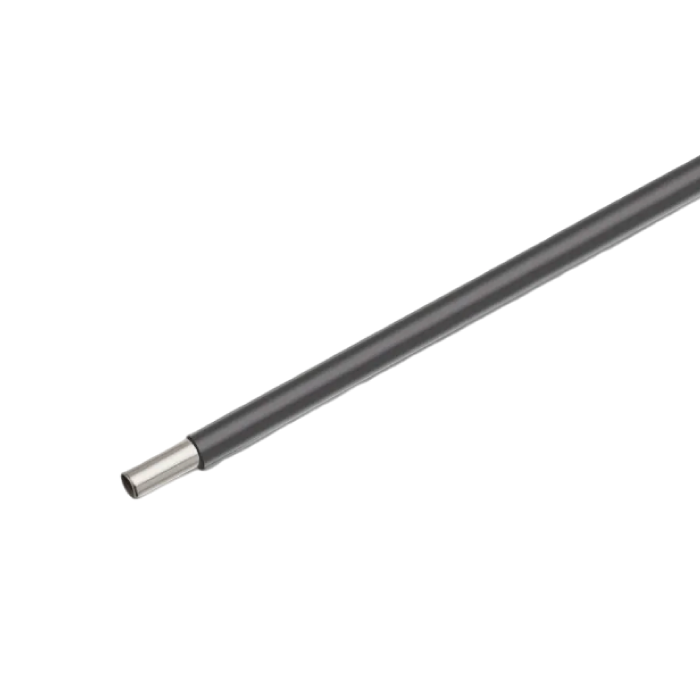
The Autolith™ Touch Probe, a 375cm-long single use device optimised for use with the SpyGlass™ DS Direct Visualization System.
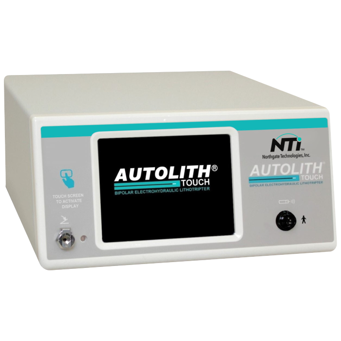
The Autolith™ Touch EHL Generator which provides a precise balance of energy to avoid probe power overload, and monitors the probe, alerting the user when it needs to be replaced.
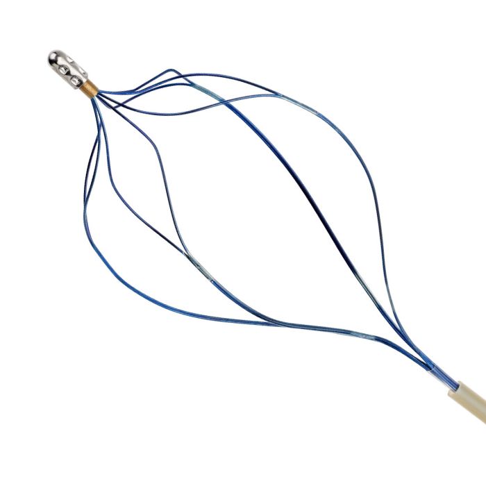
The SpyGlass™ Retrieval Basket can be used to capture and remove biliary and pancreatic stones and stone fragments visualised with the SpyGlass™ DS System.
Cholangioscopy Biopsy and Tissue Acquisition
A significant proportion (70-80%) of biliary strictures are malignant.7 Performing biopsies under direct visualisation using the SpyGlass DS System and Legacy SpyBite Biopsy Forceps enables faster, more accurate diagnosis of malignancies compared to cytology brushing.
SpyBite Max Biopsy Forceps – a design enhancement to the legacy SpyBite Biopsy Forceps – have been shown to acquire on average more than 2x tissue per bite.8-10
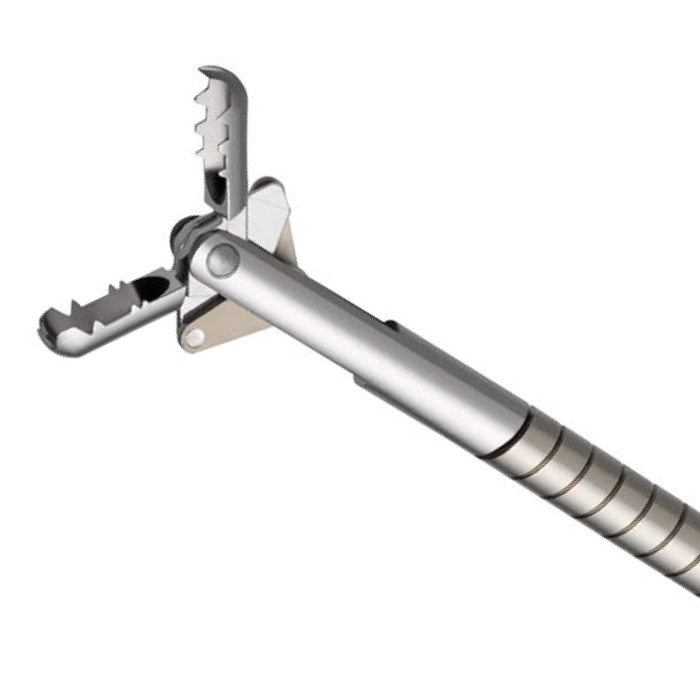
Evidence Supporting the use of SpyBite™ Max Biopsy Forceps
97%
tissue acquisition rate. Higher tissue acquisition rate may enable more efficient procedures11
89%
of physicians stated SpyBite Max acquired on average more tissue per bite than Legacy SpyBite11
88%
of physicians were more confident in their ability to acquire tissue from challenging scope positions11
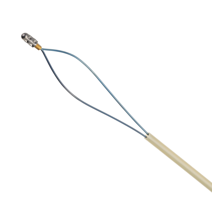
Use alongside the SpyGlass™ cholangioscopy system and the SpyGlass™ Retrieval Snare to enable efficient capture and removal of foreign bodies in the biliary and pancreatic ducts, such as migrated plastic stents, during an ERCP procedure.