")

")

")
")


")
")


Patient Screening
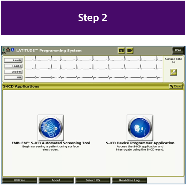
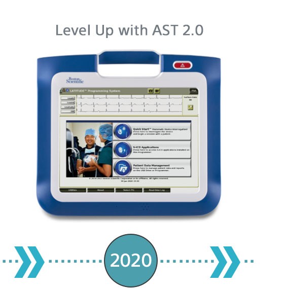
AST 2.0 Now Available through the LATITUDE™ Programming System, Model 3300
Applies the Vector Select algorithm that is used by the S-ICD to sense the cardiac signal designed to more closely represent
S-ICD device performance.1

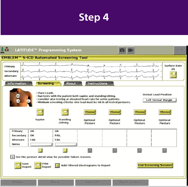
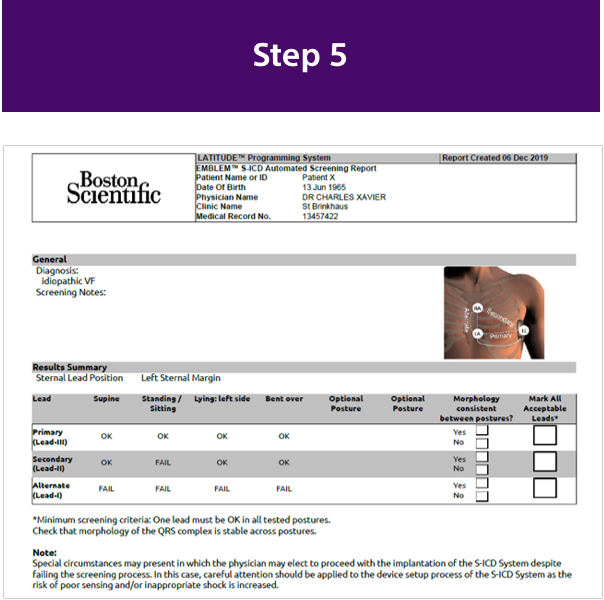
How to Do a Successful Screening
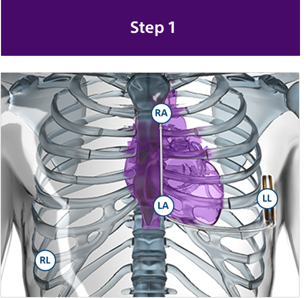
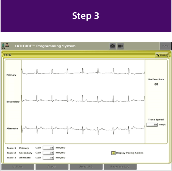
With the EMBLEM MRI S-ICD Automated Screening Tool, you can screen patients in 5 simple steps.2
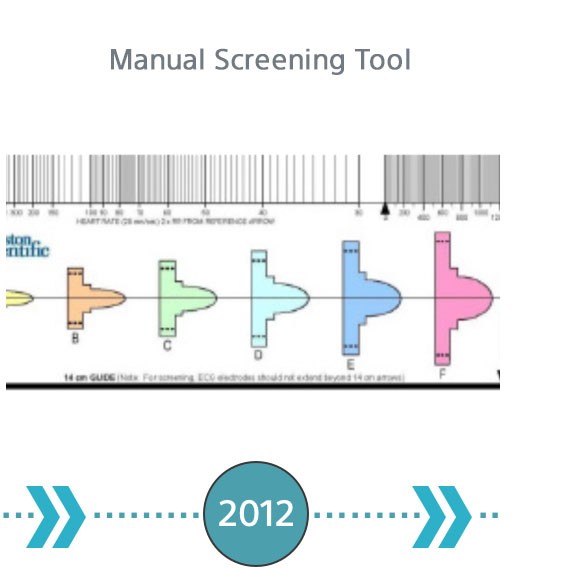
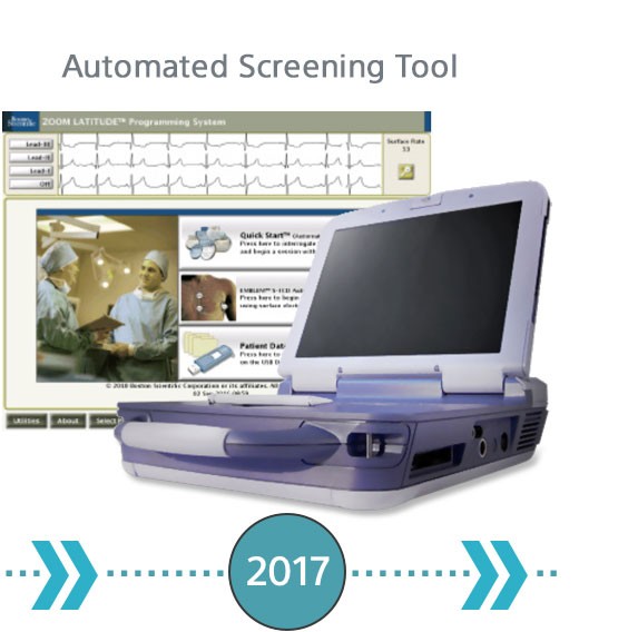
The Evolution of S-ICD Screening
Screening for S-ICD has continued to evolve to remove subjectivity while increasing efficiency and usability.
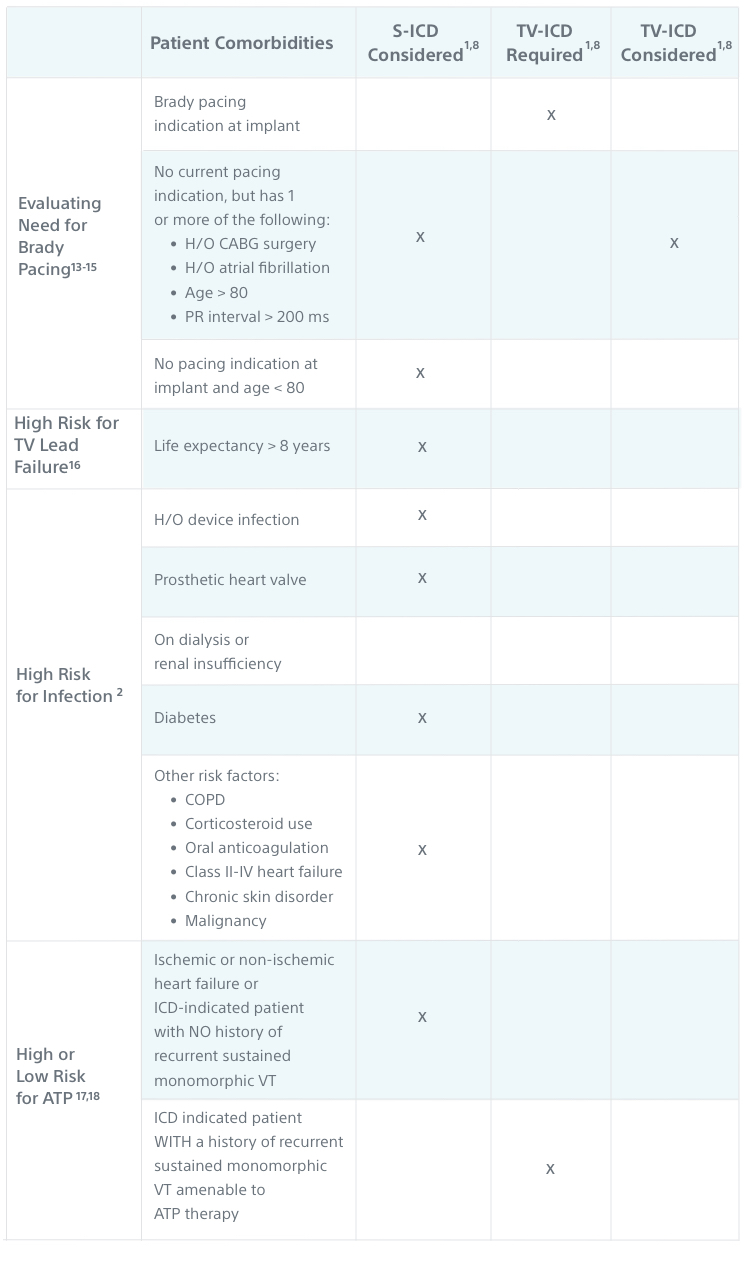
Which Patients Should Be Screened for an S-ICD?
All ICD-indicated patients without a pacing need should be considered for an S-ICD.
Patient Comorbidities |
S-ICD |
TV-ICD Required3,4 |
TV-ICD Considered3,4 |
|
|---|---|---|---|---|
| Evaluating Need for Brady Pacing5-7 | Brady pacing indication at implant |
|
|
|
No current pacing indication, but has 1 or more of the following:
|
|
|
||
| No pacing indication at implant and age < 80 |
|
|
|
|
| High Risk for TV Lead Failure8 | Life expectancy > 8 years |
|
|
|
| High Risk for Infection9 | H/O device infection |
|
|
|
| Prosthetic heart valve |
|
|
||
| On dialysis or renal insufficiency |
|
|
||
| Diabetes |
|
|
||
Other risk factors:
|
|
|
||
| High or Low Risk for ATP10,11 | Ischemic or non-ischemic heart failure or ICD-indicated patient with NO history of recurrent sustained monomorphic VT |
|
||
ICD-indicated patient WITH a history of recurrent sustained monomorphic VT amenable to ATP therapy |
|