Banner

Precise Endoscopic Resection. Confident Completion.


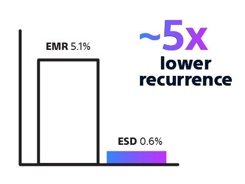
Endoscopic resection techniques are used to remove superficial gastrointestinal lesions using minimally invasive approaches. The primary techniques include EMR, ESD, and hybrid techniques, each selected based on lesion characteristics and procedural complexity.

Choosing the Right Approach

Endoscopic resection presents procedural challenges, particularly when lesion size, morphology, and location vary. These factors, along with operator expertise, can increase procedural complexity, influence workflow, and affect the likelihood of complete resection and the need for additional interventions.

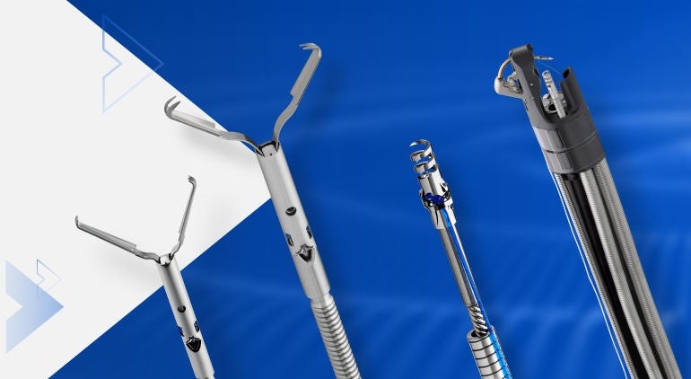
Successful resection begins with selecting an approach that matches lesion characteristics and procedural intent. Reliable resection technologies that support precise tissue capture and controlled dissection can help you manage complexity, perform consistently across a range of scenarios, and enable effective, organ-preserving treatment when clinically appropriate.
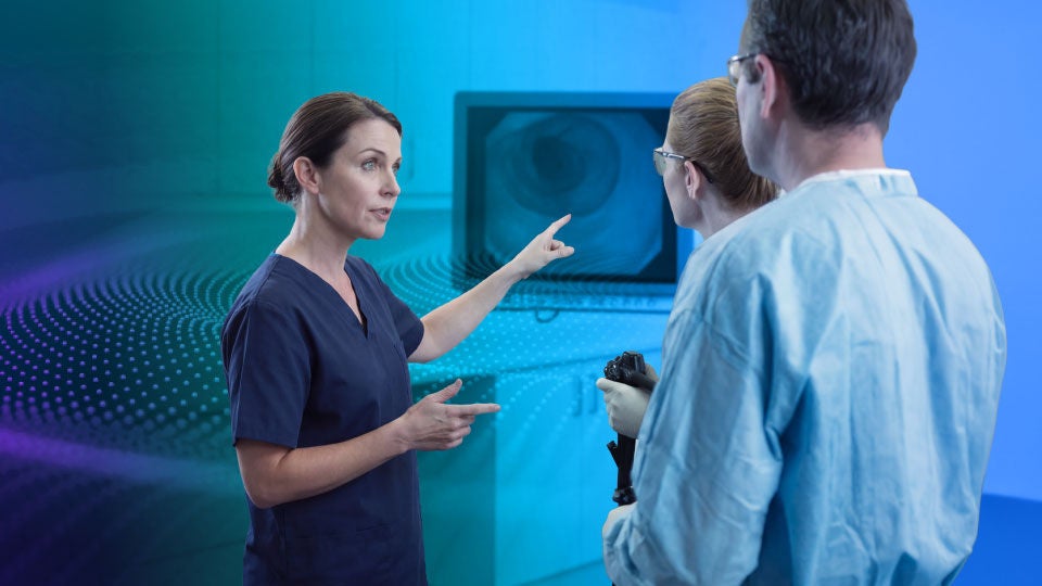
Clinical Workflow

Workflow

Our clinical workflow moves from lesion detection through technique selection (EMR, ESD, or Hybrid), to resection, and closure where required.


Our Endoscopic Resection Techniques

EMR

Endoscopic Mucosal Resection (EMR)

EMR remains an effective and efficient option for many mucosal lesions, supported by established workflows that deliver predictable and consistent outcomes.

Explore our EMR Portfolio


ESD

Endoscopic Submucosal Dissection (ESD)

ESD enables en-bloc resection where margin control and curative intent are priorities, allowing accurate histologic assessment of lateral and vertical margins.

Explore ORISE™ ProKnife


Hybrid

Hybrid Technologies

Hybrid techniques can help bridge treatment needs across lesion complexity by combining procedural efficiency with resection control, supporting physician choice and flexibility.


Expanding the Role of ESD

Doctor


ESD is increasingly used for lesions where margin control and curative intent are essential, and represents one of the more advanced endoscopic resection approaches available to endoscopists. With appropriate training and experience, a structured approach to case selection and technique can support safe integration into clinical practice.

Structured training pathways, standardised workflows, and dedicated procedural technologies support this transition, broadening access to organ-preserving treatment and enabling more physicians to manage complex lesions with confidence.


Real World Evidence

As early detection improves, more superficial GI lesions can be treated endoscopically.

Endoscopic resection techniques such as EMR and ESD are supported by a growing body of clinical evidence demonstrating strong endoscopic resection outcomes and their role in organ-preserving care.1-3

When applied in appropriately selected patients, these approaches can achieve high rates of disease eradication while reducing the need for surgical intervention.4-8

For healthcare systems, minimally invasive approaches can support efficient care delivery while maintaining strong clinical outcomes.9,10

Low rates

EMR is associated with low rates of major complications (~1.5%), which are typically manageable11

Low raecurrence

Up to ~5x lower recurrence with ESD in complex lesions12

lower cost

Endoscopic resection can reduce procedural costs compared with surgery, reflecting its less invasive nature13

Resection is Complete When Closure is Secure

Integrating closure as a planned step helps transform technical success into a confidently completed treatment pathway.

Women in Endoscopy


One Partner. Multiple Solutions.

At Boston Scientific, we deliver services and solutions designed to support professional education, operational efficiency and environmental sustainability.


Connect with us:

Connect with our team to access the support, resources, and information you need to stay informed and make the most of our endoscopy solutions.

References

1. Yang D, et al. Clin Gastroenterol Hepatol. 2018;17:1019-28.

2. Zheng X, et al. Front Med. 2025;12:1702512.

3. Moreira P, et al. J Clin Med. 2023;12(14):4777.

4. 4Akintoye, Emmanuel & Kumar, Nitin & Aihara, Hiroyuki & Nas, Hala & Thompson, Christopher. (2016). Colorectal endoscopic submucosal dissection: a systematic review and meta-analysis. Endoscopy International Open. 4. 10.1055/s-0042-114774. https://www.thiemeconnect.com/products/ejournals/pdf/10.1055/s-0042-114774.pdf Accessed September 6, 2020

5. Liu, Qing et al, Updated evaluation of endoscopic submucosal dissection versus surgery for early stage gastric cancer: a systematic review and meta-analysis, International Journal of Surgery, November 2019

6. Zhang et al. Clinical Gastroenterology and Hepatology Vol. 17, No. 1 January 2019

7. Keswani RN, Law R, Ciolino JD, et al. Adverse events after surgery for nonmalignant colon polyps are common and associated with increased length of stay and costs. Gastrointest Endosc. 2016;84(2):296-303.e1.

8. Abdelfatah MM, Barakat M, Ahmad D, et al. Long-term outcomes of endoscopic submucosal dissection versus surgery in early gastric cancer: a systematic review and meta-analysis. Eur J Gastroenterol Hepatol. 2019;31(4):418-424. doi:10.1097/MEG.0000000000001352

9. Tanaka H. Otolaryngol Online J. 2024;14(4):397.

10. Tewani S, et al. Clin Gastroenterol Hepatol. 2022;20:2680-3.

11. Pech O, et al. Gastroenterology. 2014;146(3):652-60.e1.

12. Jacques J, et al. Ann Intern Med. 2024;177(1):29-38.

13. Jacques J, et al. United European Gastroenterol J. 2019;7(1):138-45.

CAUTION: The law restricts these devices to sale by or on the order of a physician. Indications, contraindications, warnings, and instructions for use can be found in the product labelling supplied with each device or at www.IFU-BSCI.com. Products shown for INFORMATION purposes only and may not be approved or for sale in certain countries. This material not intended for use in France.