
")

")

")
")


")
")


An Innovative Approach to Liver Biopsies
 Avik Sarkar, M.D.
Assistant Professor of Medicine
Director, Bariatric Endoscopy Program
Robert Wood Johnson University Hospital
New Brunswick, NJ
 Vinod K Rustgi, M.D., MBA
Professor of Medicine
Clinical Director of Hepatology
Robert Wood Johnson University Hospital
New Brunswick, NJ
Patient History
Procedure
Specimen Preparation
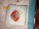
Figure 1
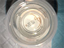
Figure 2
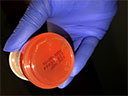
Figure 3
Outcome
Hepatologist Perspective
More Case Studies
Direct Endoscopic Visualization via Cholangioscopy Before and After Radiofrequency Ablation
Educare
To explore in-depth physician-led lectures, procedural techniques and device tutorials, visit Educare.
Get started