


")

Unusual embolization of Hip Replacement pseudotumor
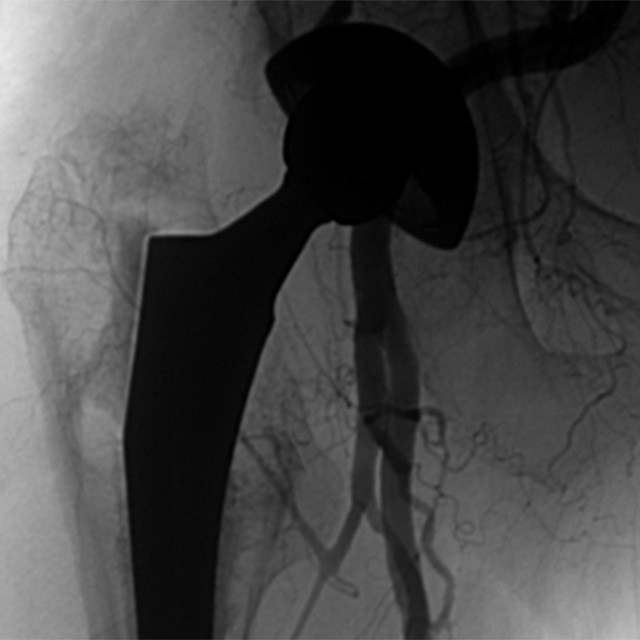
Baseline Central
An 81 year old woman, with hip replacement, was complaining of sharp pain in the prosthesis location.
The radiological examination showed periacetabular osteolysis and mass-forming tissue reaction around the metal-on-metal hip prosthesis.
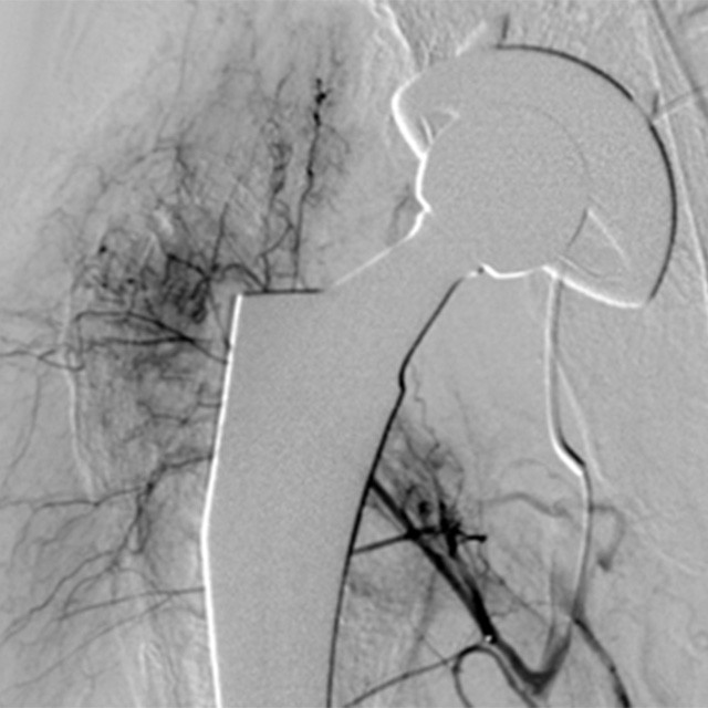
First Embolization
The angiographic examination showed some feeding vessels originating from the right femoral artery, the right iliac artery and the perforating artery.
By using a Direxion™ Hi-Flo preloaded with Fathom™-16 we catheterized the ascending branche of the lateral circumflex fermoral artery.
We embolized this vessel with 0.2cc of glue.
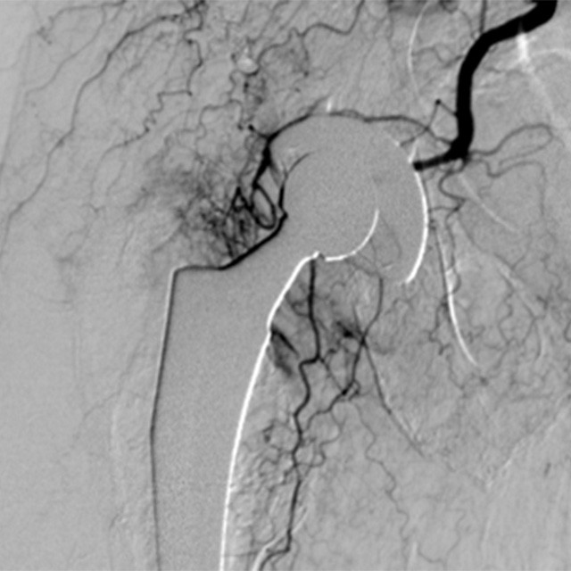
Great Control
By using the same microcatheter we cannulated the second feeding vessel originating from the femoral artery.
During the third embolization, the torque control of the Direxion™ help us to engage a small vessel originating from the iliac artery and pass through this vessel to exclude a small anastomosis to the vagina.
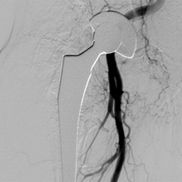
Final Result
Angiographic scan showed complete embolization of the lesion, total patency of the femoral artery and no non-target embolization.
The patient had complete pain relief and is under control.
Dott. Giuseppe Rossi – Chief of Department Angiographic Intervenional Radiology – Interventional Radiologist – Rizzoli Orthopedics Institute – Bologna
Dott. Matteo Renzulli – Interventional Radiologist – Sant’Orsola Malpighi Hospital – Bologna