


")

Selective Embolization of Traumatic Kidney Vascular Injury
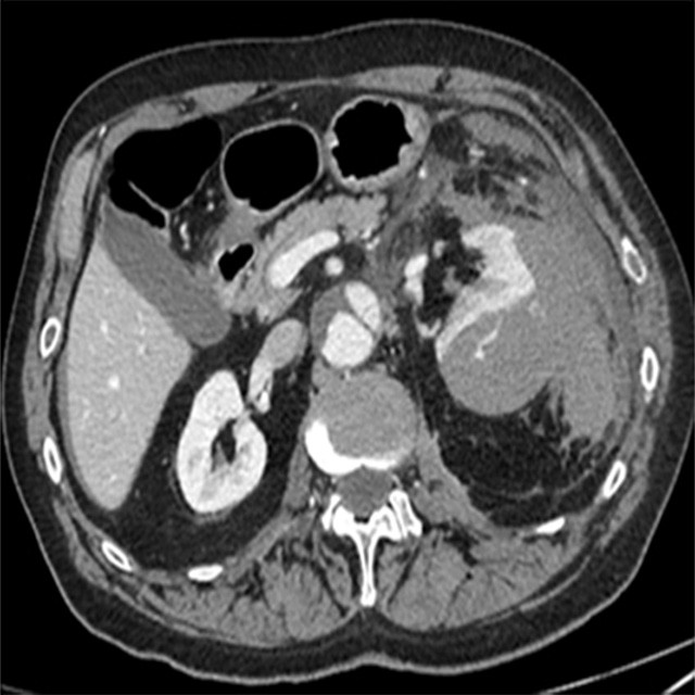
Baseline Central
A 78 year old male patient was admitted to emergency department and underwent a total body CT scan after a car accident.
The scans showed large subcapsular haematoma with active arterial supply at the middle-lower third level of the left kidney .
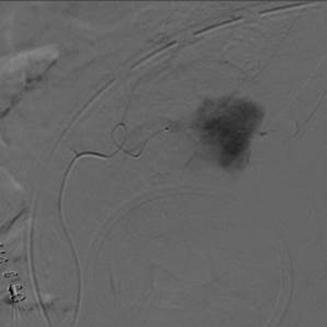
Angiography Scan
A Direxion™ 0.021" (0.53mm) microcatheter was used to engage the left renal artery and perform a super-selective catheterization of the middle-inferior lobe vessels.
The angiographic image confirmed active bleeding due to arterial laceration.
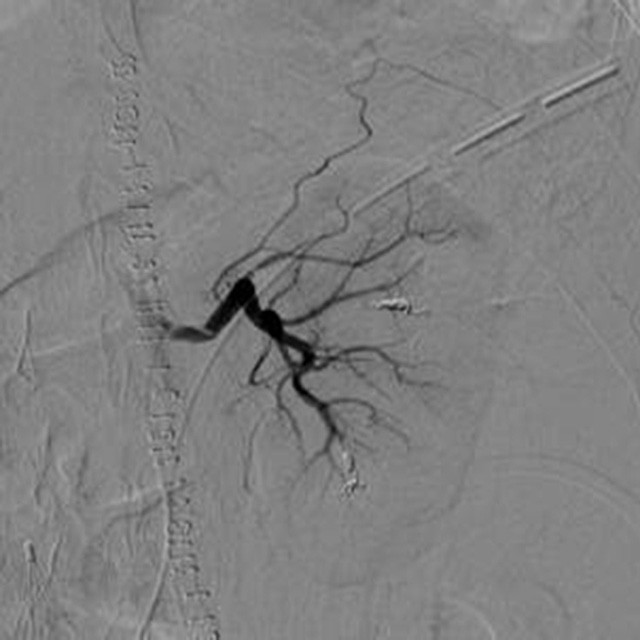
Embolization
The first embolization was performed with Interlock™ detachable coils 3x40 mm.
Then using the same Direxion™, with an accurate torquablility performance, we were able to perform distal embolization,, preserving renal parenchyma, by using Interlock™ detachable coil 2x40mm.
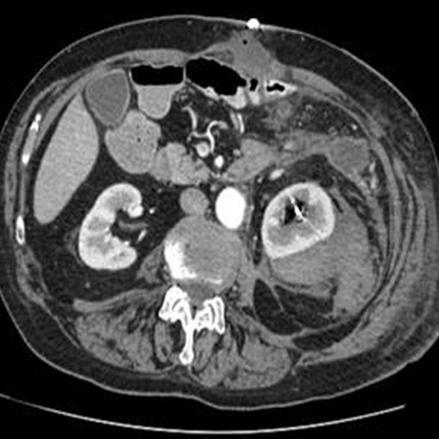
Result
The final angiographic image confirmed a very good and precise embolization.
After 5 days CT-scan showed a capsular hematoma reduction and the absence of active bleeding.
Dott. Angelo Spinazzola – Chief of Interventional Radiology Department and Interventional Radiologist – Ospedale Maggiore – Crema
Dott. Nicola Cionfoli – Interventional Radiologist - Ospedale Maggiore – Crema