


")

Retrograde revascularization of superior mesenteric artery
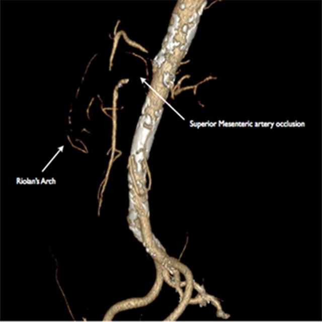
CT Scan
A 77 year old female patient with “claudicatio abdominis”, was admitted to the Interventional Radiology unit after a CT scan that showed an occlusion in the proximal segment of superior mesenteric artery (SMA) with a pseudoaneurysm at its origin.
CT scan, also showed a severe stenosis of the celiac trunk.
First Treatment
Direct revascularization of SMA was considered too risky, as the guide could cause rupture of the pseudoaneurysm.
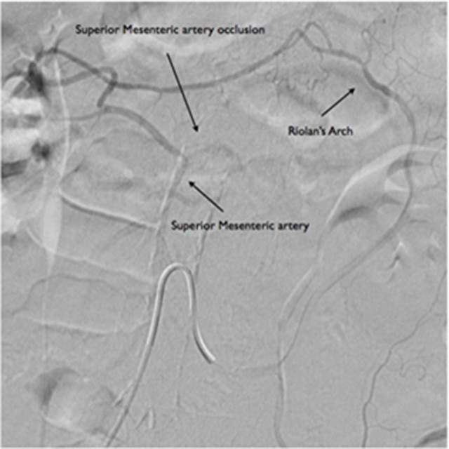
After systemic heparinization with 5000 IU, a preliminary angiography of the inferior mesenteric artery (IMA) was performed, using a 5F (1.67mm) hydrophilic catheter SIM 1 type, showing the patency of Riolano’s arch.
A Direxion™ Bern-shape microcatether, was coaxially introduced and easily pushed through the Riolano’s arch. It succesfully crossed the SMA occlusion and was advanced to the abdominal aorta.
Second Treatment
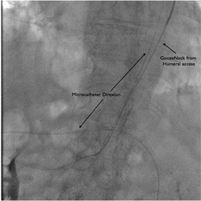
From a left humeral access, a 260 cm guidewire was captured using a Goose Neck .
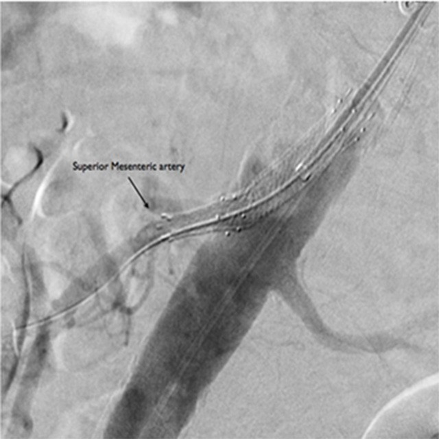
From the same access, a 7Fr (2.33mm) x 90 cm introducer sheath was advanced across the occlusion in SMA, where a covered stent was placed.
The unique Direxion nitinol design gave unrivaled steerability.
Slotted nitinol hypotube combined with braided inner liner provided an amazing pushability which helped Direxion to track onto the wire and overcame resistance encountered in complex anatomies and challenging case like this one.
Result
The final angiogram showed the complete revascularization of the branch.
No periprocedural complications occurred and the patient was discharged on the second day.
The patient had completely resolved the “claudicatio abdominis” 1 month after the procedure and a CT scan showed the patency of the stent in SMA.
Dr. Tito Torri – Chief of Radiology Department & Interventional Radiologist – "NOA Hospital" – Massa
Dr. Claudio Ceccherini - Interventional Radiologist – «NOA Hospital" – Massa