


")

Partial splenic artery embolization in the treatment of Hypersplenism
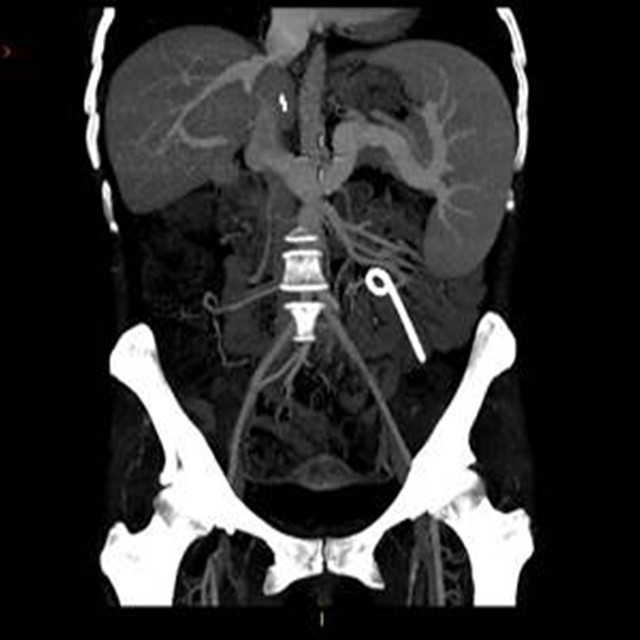
Baseline Central
A 30-year old woman with an history of cirrhosis HBV related.
Clinical history reported an HCC treated with TACE in 2013 followed by liver transplantation in August 2014.
After surgery the patient developed portal hypertension and hypersplenism.
Treatment proposed: partial splenic embolization.
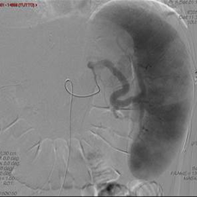
Anatomy Study
With a right femoral access, using 5F (1.67mm) sheath and 4F (1.33mm) cobra catheter, we catheterized the splenic artery to perform a diagnostic angiography.
Thereafter we decided to go distally to the peripheral intrasplenic branches to perform a more selective embolization.
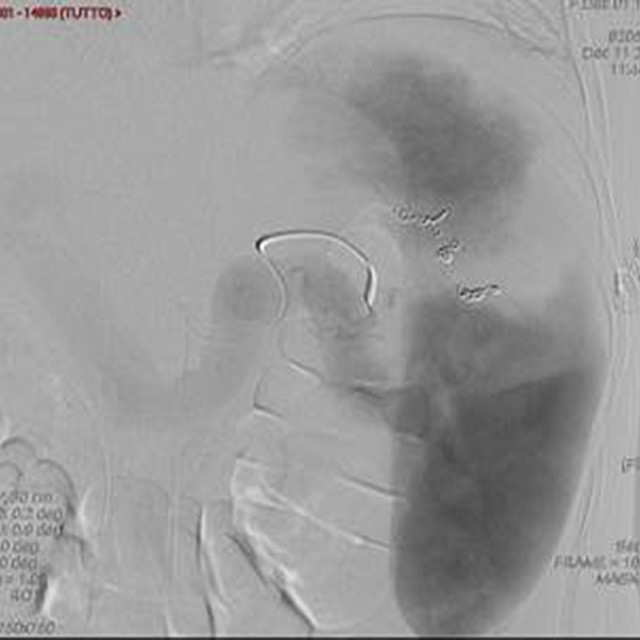
Superselective Treatment
Subsequently we embolized those branches using microcoils. After embolization, the patient developed transient abdominal pain, remitted with pain relief drugs.
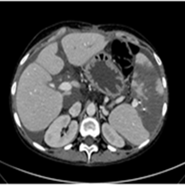
Result
Post-embolization CT-scan demonstrated a successful embolization with evidence of partial devascularization of the spleen, with minimal perisplenic and perihepatic fluid, and the absence of major complications.
The patient showed significant reduction of portal pressure and was discharged some days after. She is still in follow-up at our institution in good clinical conditions.
Dr. Rita Golfieri – Chief of Radiology Unit and Vice Director of Department of Digestive Disease – Sant’Orsola Malpighi Hospital - Bologna
Dr. Francesco Modestino – Interventional Radiologist – Sant’Orsola Malpighi Hospital - Bologna