


")

Embolization of Aortic Aneurysm sac tipe II endoleak
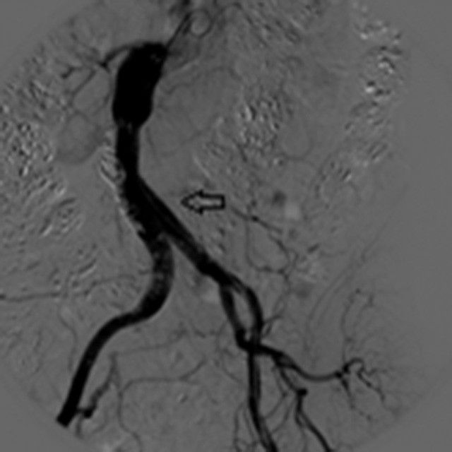
Baseline Central
A 59 year old woman with an history of fusiform AAA treated with EVAR and previous coil placement in the false lumen for type II endoleak.
CT angiography scan showed an aneurysm sac enlargement and type IIb endoleak.
The angiographic evaluation showed two small branch vessels filling the aneurysm sac.
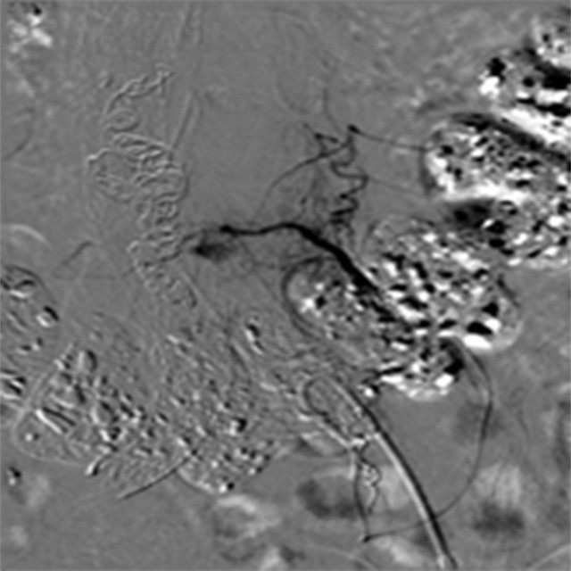
First Embolization
A Bern Shape Direxion™ Torquable Microcatheter and a Fathom-16 guidewire were used to select distally those tiny branches.
Microcoils were deployed and glue was injected.
Angiography of the SMA depicted a long tortuous Arch of Riolan and a smooth blush within the aneurysm sac.
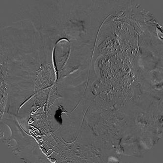
A Challenging Anatomy
The same Direxion™ was used to cannulate the middle colic artery, the Arch of Riolan, surpassing winding loops and a tight turn at the left colic flexure, and the left colic artery.
The tip of the microcatheter was advanced to the arterial ostium of the inferior mesenteric artery and into the lumen of the sac without rejecting the guiding catheter.
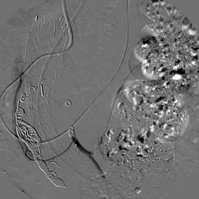
Result
Dott. Paolo Faccioli – Chief of Interventional Radiology Department – A.Manzoni Hospital – Lecco
Dott. Simone Limonta – Interventional Radiologist – A.Manzoni Hospital – Lecco