")

")

")
")


")
")


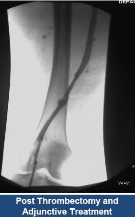
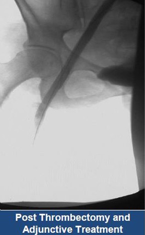
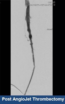
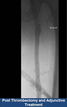
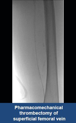
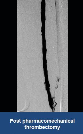
AngioJet™
Ultra Thrombectomy System
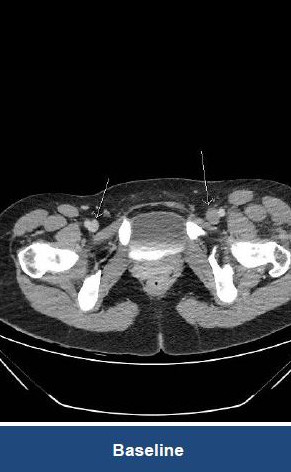
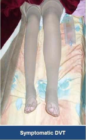
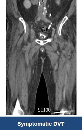
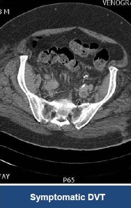
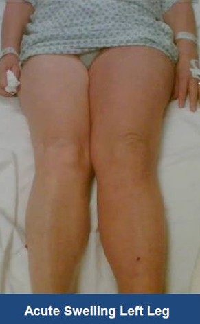
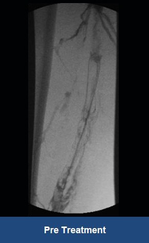
Thrombectomy of Massive DVT

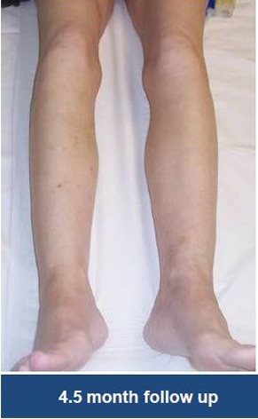
Thrombectomy of Bilateral DVT
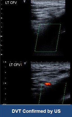
Thrombectomy of DVT
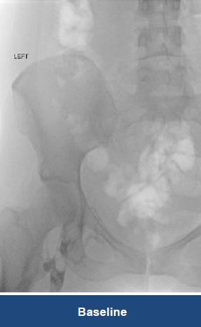
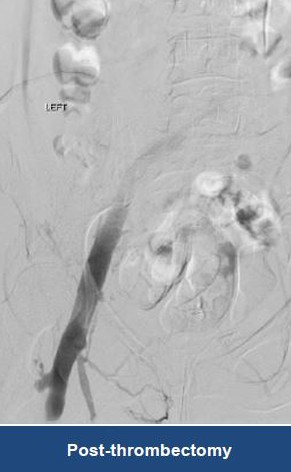
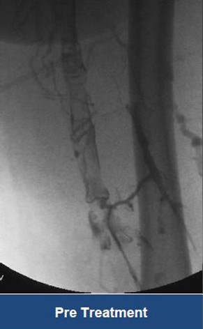
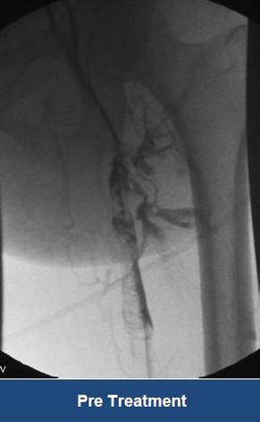
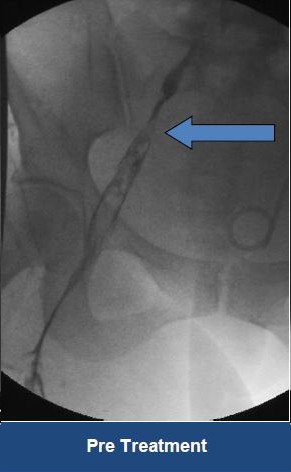
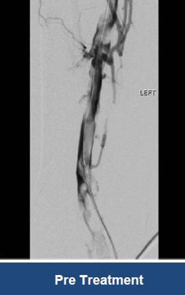
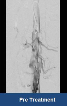
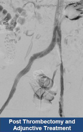
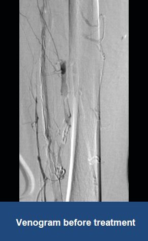
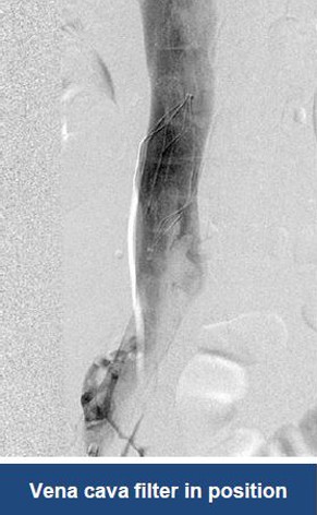
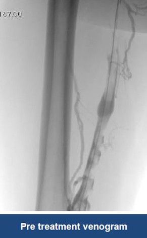
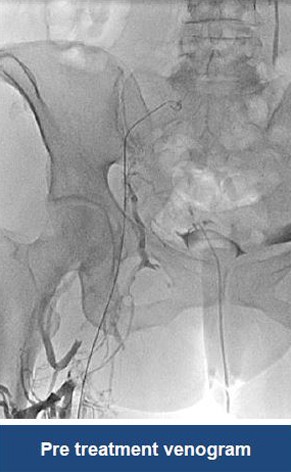
Treatment plan:
- Left popliteal access
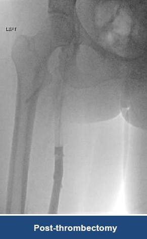
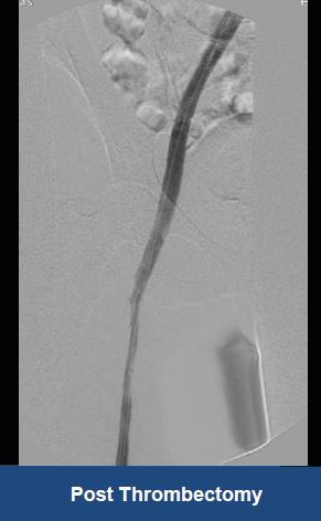
- AngioJet Thrombectomy on affected areas
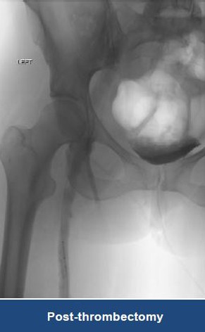
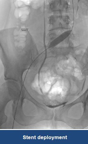
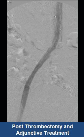
- Stent May Thurner lesion

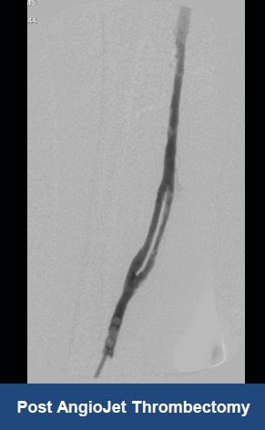

Thrombectomy of DVT
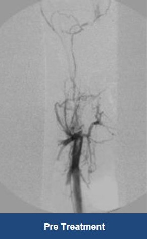
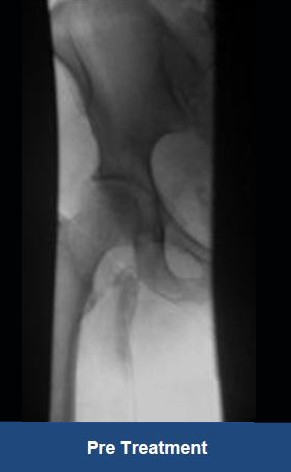
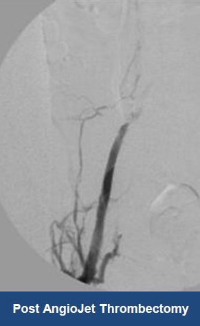
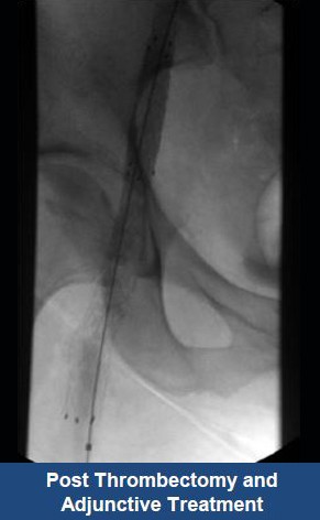
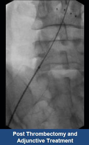
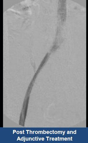
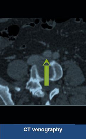
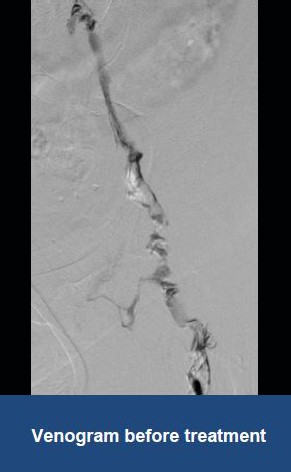
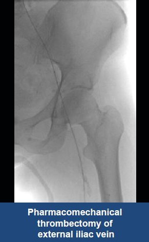
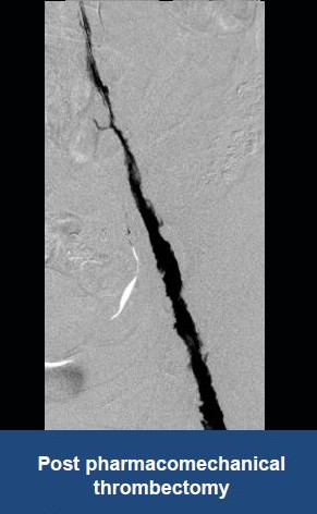
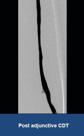

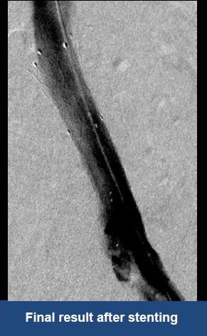
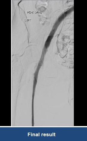
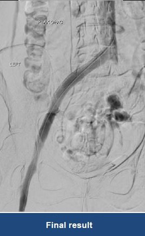
Thrombectomy of Massive Iliofemoral DVT and Stenting for May-Thurner Syndrome
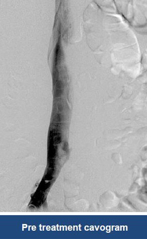
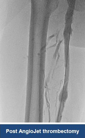
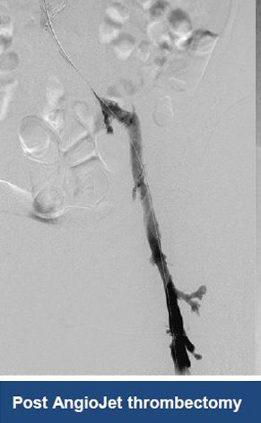
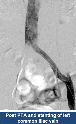
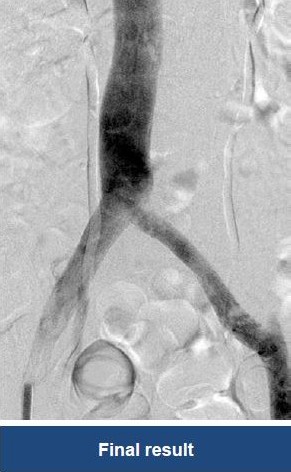
Acute Left leg Ilio femoral DVT managed with AngioJet™ Thrombectomy System and Wallstent Uni™ endoprothesis