
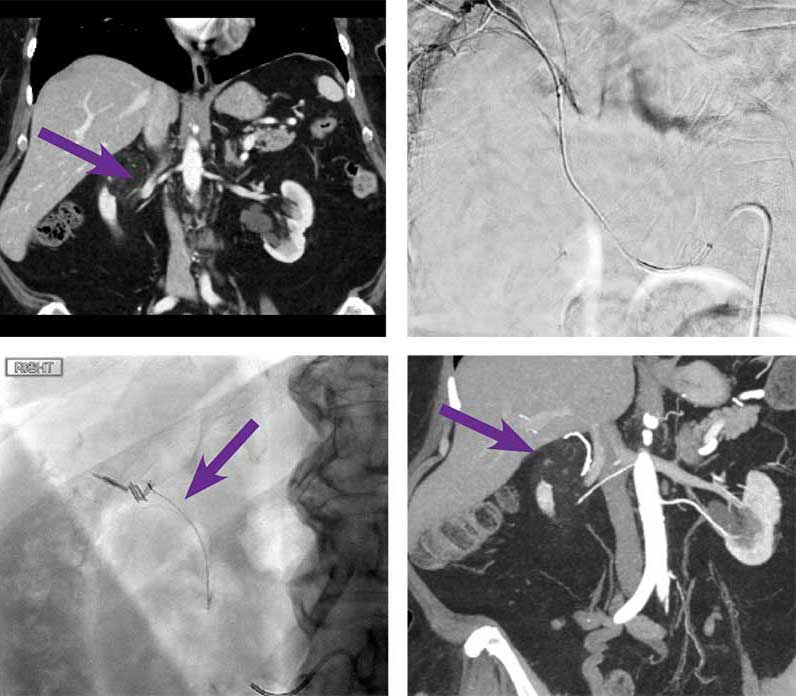
Upper pole angiomyolipoma (AML) embolization
Courtesy of Dr. Mikin Patel I University of Chicago
Presentation
75-year-old female with right upper pole AML. Lesion was incidentally found when scanning for generalized abdominal pain.
Intervention used
Using a 5F SIM1 catheter, the right renal artery was selected, and angiogram was performed. There was a small vertically oriented artery arising from a superior segmental arterial branch that courses superior to the renal parenchyma in the expected location of the angiomyolipoma seen on recent CT. Using a RenegadeTM STC Microcatheter, the small vertically oriented artery that courses beyond the renal parenchyma was selected and selective angiogram was performed. No renal parenchymal staining was seen. No arterial venous shunting was also observed. 0.2 cc of Obsidio Embolic deployed into 1 mm diameter vessel. Post embolization angiogram showed nonopacification of the occluded this vessel with sparing of the remaining renal vessels and good perfusion to the remainder of the kidney.
Outcome
Repeat CT at 6 weeks demonstrating interval decrease in size of the AML from 6.2 cm to 5.2 cm.
Results from case studies are not necessarily predictive of results in other cases. Results in other cases may vary.