Clean margins support accurate diagnosis
Optimal Tissue Samples Help Support Accurate Diagnoses:
Medical Director at Boston Scientific
There are two key Elements to Optimal Biopsy Samples
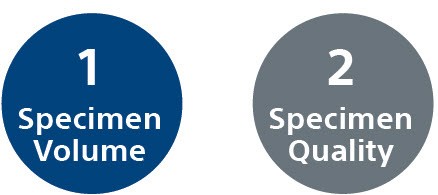
Specimen Volume and Quality
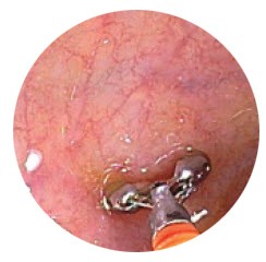
The "UPPER" Technique1
- Use largest size forceps that will fit in the biopsy channel of the scope
- Position forceps en face to the mucosa ("turn-in" technique versus "scissor" technique versus scope rotation)
- Partially collapse lumen (reduce wall tension)
- Energize the bite (firm, snap the forceps closed, short throw/pull into scope)
- Repeat biopsies - especially in small and large intestine (large forceps aids in size of second biopsy sample)
Handling Techniques to Improve Specimen Quality
- Identify "inadequate" samples
- Inadequate samples (size and quality) compromise the quality of the overall specimen - Optimal extraction technique -
- Use blunt probe to push biopsy out of the base of the opened forceps cups
- Specimen can be squashed if pushed from top of open forceps - Shaking biopsies off forceps into fixative can traumatize tissue and cause erosion of the epithelium
- Try to keep to a maximum of 4 biopsies/fixative jar
Driving Quality through Communication
| Lesion Description | Use simple language, give visuals, e.g. “thick folds” vs “hypertrophic” |
|---|---|
| Biopsy Instrument | Provide detail on device, e.g. “hot biopsy forceps” vs. “pinch forceps” |
| For Polyps | Give size and style (sessile or pedunculated) |
| Key Drugs | Provide inclusive and detailed list of medications |
| History | Provide brief 1-2 line history |
| Questions for Pathologist | Be specific about what questions you want answered |
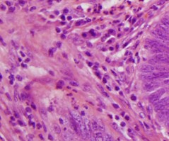
Reduce Crush Artifact
- Crush artifact is the tearing or compression of tissue often caused by the penetration of the forceps during biopsy
- Every biopsy has some crush artifact, typically at the periphery.
- Crushed tissue is difficult to interpret by a pathologist and can impact the ability to make definitive diagnoses
- Minimize the amount of tissue crushed by:
– Minimizing the handling of the specimen
– Not shaking the biopsy off the forceps
– Not pushing the biopsy out of the cup from the top
Disease States
GI disease states are complex and require specialized expertise and partnership between clinical staff and the pathology provider
The likelihood of an accurate diagnosis may increase with:
- Selection of proper tools with adequate tissue sampling
- Tailored approaches to specific disease states
- Proper specimen handling
- Detailed communication between staff and pathology lab
- Expert, specialized pathologists