
")

")

")
")


")
")


Rotablator™ Case Study: Facilitating Stent Delivery
Rotational Atherectomy System
Diagnosis
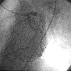
- Baseline angiography showed a small, non-dominate right coronary artery
- The left anterior descending presented mild disease
- The distal left circumflex showed 90% stenosis
- The left ventricle function was normal

Procedure
Treatment of Circumflex
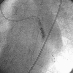
- Placed a guide catheter to facilitate potential use of various interventional devices
- A guidewire was used to cross the distal left circumflex lesion
- Unsuccessful dilatation of the lesion attempted with a 4.0 mm × 20 mm balloon catheter
- Unsuccessful dilatation of the lesion attempted with 4.0 mm × 15 mm non-compliant balloon catheter at 14 ATM
- A 4.0 mm × 20 mm non-compliant balloon at 14 ATM also proved unsuccessful in expanding the lesion
- A 4.0 mm × 10 mm Cutting Balloon™ Device was deployed, but expansion of the left circumflex proved unsuccessful

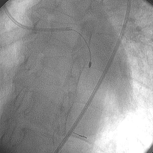
Post-ROTABLATOR System
The medical team decided to use the Rotablator System. A 1.75 mm burr was passed through the lesion without difficulty over the RotaWire™ Floppy Guide Wire.
- Following the use of the Rotablator System, the lesion was post dilated with a 4.0 mm × 20 mm PTCA balloon catheter
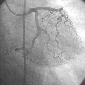
- The use of the Rotablator System facilitated the delivery and expansion of a 4.0 mm × 9.0 mm stent with good angiographic appearance


Physician Commentary
The Rotablator System served as a facilitator of lesion compliance and ultimately, stent delivery and expansion.
This case illustrates the ability of the Rotablator System to modify calcified atherosclerotic plaque in order to facilitate optimal stent delivery and expansion. Angiographic assessment did not uncover the calcified nature of the lesion.
After multiple balloon dilatation attempts with non-compliant balloons, the level and extent of calcification was made apparent. Although the 1.75 mm burr did not increase the lumen size, it did allow for subsequent expansion of the lesion by means of a PTCA balloon catheter and stent.